• 文献检索
  • 文档翻译
  • 深度研究
  • 学术资讯
  • Suppr Zotero 插件Zotero 插件
  • 邀请有礼
  • 套餐&价格
  • 历史记录
应用&插件
Suppr Zotero 插件Zotero 插件浏览器插件Mac 客户端Windows 客户端微信小程序
定价
高级版会员购买积分包购买API积分包
服务
文献检索文档翻译深度研究API 文档MCP 服务
关于我们
关于 Suppr公司介绍联系我们用户协议隐私条款
关注我们

Suppr 超能文献

核心技术专利:CN118964589B侵权必究
粤ICP备2023148730 号-1Suppr @ 2026

文献检索

告别复杂PubMed语法,用中文像聊天一样搜索,搜遍4000万医学文献。AI智能推荐,让科研检索更轻松。

立即免费搜索

文件翻译

保留排版,准确专业,支持PDF/Word/PPT等文件格式,支持 12+语言互译。

免费翻译文档

深度研究

AI帮你快速写综述,25分钟生成高质量综述,智能提取关键信息,辅助科研写作。

立即免费体验

三维打印在心胸外科的应用

Cardiothoracic Applications of 3-dimensional Printing.

作者信息

Giannopoulos Andreas A, Steigner Michael L, George Elizabeth, Barile Maria, Hunsaker Andetta R, Rybicki Frank J, Mitsouras Dimitris

机构信息

*Applied Imaging Science Laboratory †Radiology Department, Division of Thoracic Imaging, Brigham and Women's Hospital, Boston, MA ‡Department of Radiology, The University of Ottawa Faculty of Medicine and The Ottawa Hospital Research Institute, Ottawa, ON, Canada.

出版信息

J Thorac Imaging. 2016 Sep;31(5):253-72. doi: 10.1097/RTI.0000000000000217.

DOI:10.1097/RTI.0000000000000217
PMID:27149367
原文链接:https://pmc.ncbi.nlm.nih.gov/articles/PMC4993676/
Abstract

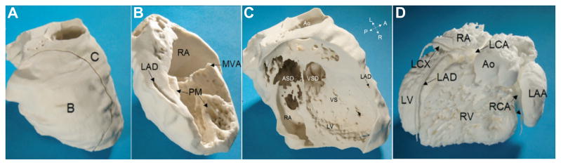
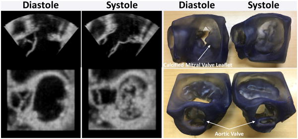
Medical 3-dimensional (3D) printing is emerging as a clinically relevant imaging tool in directing preoperative and intraoperative planning in many surgical specialties and will therefore likely lead to interdisciplinary collaboration between engineers, radiologists, and surgeons. Data from standard imaging modalities such as computed tomography, magnetic resonance imaging, echocardiography, and rotational angiography can be used to fabricate life-sized models of human anatomy and pathology, as well as patient-specific implants and surgical guides. Cardiovascular 3D-printed models can improve diagnosis and allow for advanced preoperative planning. The majority of applications reported involve congenital heart diseases and valvular and great vessels pathologies. Printed models are suitable for planning both surgical and minimally invasive procedures. Added value has been reported toward improving outcomes, minimizing perioperative risk, and developing new procedures such as transcatheter mitral valve replacements. Similarly, thoracic surgeons are using 3D printing to assess invasion of vital structures by tumors and to assist in diagnosis and treatment of upper and lower airway diseases. Anatomic models enable surgeons to assimilate information more quickly than image review, choose the optimal surgical approach, and achieve surgery in a shorter time. Patient-specific 3D-printed implants are beginning to appear and may have significant impact on cosmetic and life-saving procedures in the future. In summary, cardiothoracic 3D printing is rapidly evolving and may be a potential game-changer for surgeons. The imager who is equipped with the tools to apply this new imaging science to cardiothoracic care is thus ideally positioned to innovate in this new emerging imaging modality.

摘要

医学三维(3D)打印正在成为一种在许多外科专业中指导术前和术中规划的临床相关成像工具,因此可能会促成工程师、放射科医生和外科医生之间的跨学科合作。来自计算机断层扫描、磁共振成像、超声心动图和旋转血管造影等标准成像模态的数据可用于制作人体解剖结构和病理的实物大小模型,以及针对患者的植入物和手术导板。心血管3D打印模型可改善诊断并有助于进行高级术前规划。报告的大多数应用涉及先天性心脏病以及瓣膜和大血管病变。打印模型适用于规划手术和微创手术。据报道,其在改善治疗效果、将围手术期风险降至最低以及开发诸如经导管二尖瓣置换等新手术方面具有附加价值。同样,胸外科医生正在使用3D打印来评估肿瘤对重要结构的侵犯,并协助诊断和治疗上下气道疾病。解剖模型使外科医生能够比查看图像更快地吸收信息,选择最佳手术方法,并在更短时间内完成手术。针对患者的3D打印植入物开始出现,未来可能会对美容和挽救生命的手术产生重大影响。总之,心胸外科3D打印正在迅速发展,可能会成为外科医生的一个潜在变革因素。因此,配备有将这种新成像科学应用于心胸护理工具的成像专家处于在这种新兴成像模态中进行创新的理想位置。

https://cdn.ncbi.nlm.nih.gov/pmc/blobs/6f4b/4993676/3ad349a425ee/nihms775999f17.jpg
https://cdn.ncbi.nlm.nih.gov/pmc/blobs/6f4b/4993676/6e8310c2286e/nihms775999f1.jpg
https://cdn.ncbi.nlm.nih.gov/pmc/blobs/6f4b/4993676/84052d19bdbc/nihms775999f2.jpg
https://cdn.ncbi.nlm.nih.gov/pmc/blobs/6f4b/4993676/891d66bcbd55/nihms775999f3.jpg
https://cdn.ncbi.nlm.nih.gov/pmc/blobs/6f4b/4993676/0af50b469b4e/nihms775999f4.jpg
https://cdn.ncbi.nlm.nih.gov/pmc/blobs/6f4b/4993676/0e0a96c2e77c/nihms775999f5.jpg
https://cdn.ncbi.nlm.nih.gov/pmc/blobs/6f4b/4993676/1c45ced06fcb/nihms775999f6.jpg
https://cdn.ncbi.nlm.nih.gov/pmc/blobs/6f4b/4993676/85021dfdf86f/nihms775999f7.jpg
https://cdn.ncbi.nlm.nih.gov/pmc/blobs/6f4b/4993676/c4f56a6fad76/nihms775999f8.jpg
https://cdn.ncbi.nlm.nih.gov/pmc/blobs/6f4b/4993676/f57a10f526b6/nihms775999f9.jpg
https://cdn.ncbi.nlm.nih.gov/pmc/blobs/6f4b/4993676/d3be4c185fb1/nihms775999f10.jpg
https://cdn.ncbi.nlm.nih.gov/pmc/blobs/6f4b/4993676/63689dc4b19c/nihms775999f11.jpg
https://cdn.ncbi.nlm.nih.gov/pmc/blobs/6f4b/4993676/2706560a4629/nihms775999f12.jpg
https://cdn.ncbi.nlm.nih.gov/pmc/blobs/6f4b/4993676/f9327c847948/nihms775999f13.jpg
https://cdn.ncbi.nlm.nih.gov/pmc/blobs/6f4b/4993676/2196d4a03064/nihms775999f14.jpg
https://cdn.ncbi.nlm.nih.gov/pmc/blobs/6f4b/4993676/67e246795ef5/nihms775999f15.jpg
https://cdn.ncbi.nlm.nih.gov/pmc/blobs/6f4b/4993676/3a9a13918db7/nihms775999f16.jpg
https://cdn.ncbi.nlm.nih.gov/pmc/blobs/6f4b/4993676/3ad349a425ee/nihms775999f17.jpg

相似文献

1
Cardiothoracic Applications of 3-dimensional Printing.三维打印在心胸外科的应用
J Thorac Imaging. 2016 Sep;31(5):253-72. doi: 10.1097/RTI.0000000000000217.
2
Hybrid 3D printing: a game-changer in personalized cardiac medicine?混合3D打印:个性化心脏医学的变革者?
Expert Rev Cardiovasc Ther. 2015 Dec;13(12):1281-4. doi: 10.1586/14779072.2015.1100076. Epub 2015 Oct 14.
3
Innovations in Preoperative Planning: Insights into Another Dimension Using 3D Printing for Cardiac Disease.术前规划的创新:利用3D打印技术对心脏病进行另一维度的洞察。
J Cardiothorac Vasc Anesth. 2018 Aug;32(4):1937-1945. doi: 10.1053/j.jvca.2017.11.037. Epub 2017 Nov 23.
4
Patient-specific three-dimensional printing for Kommerell's diverticulum.基于患者个体的 Kommerell 憩室三维打印。
Int J Cardiol. 2018 Mar 15;255:184-187. doi: 10.1016/j.ijcard.2017.12.065. Epub 2017 Dec 26.
5
Preoperative Planning Using 3D Printing Technology in Orthopedic Surgery.骨科手术中使用3D打印技术的术前规划
Biomed Res Int. 2021 Oct 12;2021:7940242. doi: 10.1155/2021/7940242. eCollection 2021.
6
Three-dimensional printing and virtual surgery for congenital heart procedural planning.三维打印和虚拟手术在先天性心脏病治疗规划中的应用。
Birth Defects Res. 2018 Aug 1;110(13):1082-1090. doi: 10.1002/bdr2.1370. Epub 2018 Aug 6.
7
The Various Applications of 3D Printing in Cardiovascular Diseases.3D 打印在心血管疾病中的各种应用。
Curr Cardiol Rep. 2018 May 10;20(6):47. doi: 10.1007/s11886-018-0992-9.
8
Application of 3-Dimensional Printing in a Case of Osteogenesis Imperfecta for Patient Education, Anatomic Understanding, Preoperative Planning, and Intraoperative Evaluation.三维打印在一例成骨不全症患者中的应用:用于患者教育、解剖学理解、术前规划及术中评估
World Neurosurg. 2017 Nov;107:1049.e1-1049.e7. doi: 10.1016/j.wneu.2017.08.026. Epub 2017 Aug 18.
9
3D printing for preoperative planning and surgical training: a review.用于术前规划和手术训练的3D打印:综述
Biomed Microdevices. 2018 Aug 4;20(3):65. doi: 10.1007/s10544-018-0301-9.
10
[Three-dimensional virtual and printed models improve preoperative planning and promote patient-safety in complex congenital and pediatric cardiac surgery].三维虚拟模型和打印模型改善复杂先天性和小儿心脏手术的术前规划并提高患者安全性
Orv Hetil. 2019 May;160(19):747-755. doi: 10.1556/650.2019.31393.

引用本文的文献

1
Optimization of Fast Non-Local Means Noise Reduction Algorithm Parameter in Computed Tomographic Phantom Images Using 3D Printing Technology.利用3D打印技术对计算机断层扫描体模图像中快速非局部均值降噪算法参数进行优化
Diagnostics (Basel). 2024 Jul 23;14(15):1589. doi: 10.3390/diagnostics14151589.
2
Sculpting the future: A narrative review of 3D printing in plastic surgery and prosthetic devices.塑造未来:整形手术和假肢装置中3D打印的叙述性综述
Health Sci Rep. 2024 Jun 23;7(6):e2205. doi: 10.1002/hsr2.2205. eCollection 2024 Jun.
3
Treatment of complex limb fractures with 3D printing technology combined with personalized plates: a retrospective study of case series and literature review.

本文引用的文献

1
3D printed ventricular septal defect patch: a primer for the 2015 Radiological Society of North America (RSNA) hands-on course in 3D printing.3D打印室间隔缺损补片:2015年北美放射学会(RSNA)3D打印实践课程入门指南
3D Print Med. 2015;1(1):3. doi: 10.1186/s41205-015-0002-4. Epub 2015 Nov 27.
2
The residual STL volume as a metric to evaluate accuracy and reproducibility of anatomic models for 3D printing: application in the validation of 3D-printable models of maxillofacial bone from reduced radiation dose CT images.残余STL体积作为评估3D打印解剖模型准确性和可重复性的指标:在低辐射剂量CT图像的颌面骨3D可打印模型验证中的应用
3D Print Med. 2015;1(1):2. doi: 10.1186/s41205-015-0003-3. Epub 2015 Nov 27.
3
3D打印技术联合个性化接骨板治疗复杂四肢骨折:病例系列回顾性研究及文献综述
Front Surg. 2024 May 16;11:1383401. doi: 10.3389/fsurg.2024.1383401. eCollection 2024.
4
Advancing Surgical Arrhythmia Ablation: Novel Insights on 3D Printing Applications and Two Biocompatible Materials.推进手术性心律失常消融:关于3D打印应用和两种生物相容性材料的新见解
Biomedicines. 2024 Apr 15;12(4):869. doi: 10.3390/biomedicines12040869.
5
Three-dimensional models for coronary artery fistulas: to print, or not to print-that is the question.冠状动脉瘘的三维模型:打印还是不打印——这是个问题。
Eur Heart J Case Rep. 2024 Feb 3;8(2):ytae069. doi: 10.1093/ehjcr/ytae069. eCollection 2024 Feb.
6
Cardiovascular Computed Tomography in the Diagnosis of Cardiovascular Disease: Beyond Lumen Assessment.心血管计算机断层扫描在心血管疾病诊断中的应用:超越管腔评估
J Cardiovasc Dev Dis. 2024 Jan 12;11(1):22. doi: 10.3390/jcdd11010022.
7
The Quantitative Impact of Using 3D Printed Anatomical Models for Surgical Planning Optimization: Literature Review.使用3D打印解剖模型优化手术规划的定量影响:文献综述
3D Print Addit Manuf. 2023 Oct 1;10(5):1130-1139. doi: 10.1089/3dp.2021.0188. Epub 2023 Oct 10.
8
Three-Dimensional Bioprinting in Cardiovascular Disease: Current Status and Future Directions.三维生物打印在心血管疾病中的应用:现状与未来方向。
Biomolecules. 2023 Jul 28;13(8):1180. doi: 10.3390/biom13081180.
9
Trends and Challenges in the Development of 3D-Printed Heart Valves and Other Cardiac Implants: A Review of Current Advances.3D打印心脏瓣膜及其他心脏植入物发展的趋势与挑战:当前进展综述
Cureus. 2023 Aug 9;15(8):e43204. doi: 10.7759/cureus.43204. eCollection 2023 Aug.
10
Medical 3D Printing Using Desktop Inverted Vat Photopolymerization: Background, Clinical Applications, and Challenges.使用桌面倒置式光聚合的医学3D打印:背景、临床应用及挑战
Bioengineering (Basel). 2023 Jun 30;10(7):782. doi: 10.3390/bioengineering10070782.
3D Printed Cardiac Phantom for Procedural Planning of a Transcatheter Native Mitral Valve Replacement.
用于经导管原生二尖瓣置换术程序规划的3D打印心脏模型
Proc SPIE Int Soc Opt Eng. 2016 Feb 27;9789. doi: 10.1117/12.2216952. Epub 2016 Apr 5.
4
Three-dimensional printing of MRI-visible phantoms and MR image-guided therapy simulation.磁共振成像可见体模的三维打印及磁共振图像引导治疗模拟
Magn Reson Med. 2017 Feb;77(2):613-622. doi: 10.1002/mrm.26136. Epub 2016 Feb 11.
5
Treatment Planning for Image-Guided Neuro-Vascular Interventions Using Patient-Specific 3D Printed Phantoms.使用患者特异性3D打印模型进行图像引导神经血管介入治疗的治疗计划
Proc SPIE Int Soc Opt Eng. 2015 Feb 21;9417. doi: 10.1117/12.2081997. Epub 2015 Mar 19.
6
3D printing based on cardiac CT assists anatomic visualization prior to transcatheter aortic valve replacement.基于心脏CT的3D打印有助于在经导管主动脉瓣置换术前进行解剖可视化。
J Cardiovasc Comput Tomogr. 2016 Jan-Feb;10(1):28-36. doi: 10.1016/j.jcct.2015.12.004. Epub 2015 Dec 12.
7
Three-dimensional Physical Modeling: Applications and Experience at Mayo Clinic.三维物理建模:梅奥诊所的应用和经验。
Radiographics. 2015 Nov-Dec;35(7):1989-2006. doi: 10.1148/rg.2015140260.
8
Medical 3D Printing for the Radiologist.面向放射科医生的医学3D打印
Radiographics. 2015 Nov-Dec;35(7):1965-88. doi: 10.1148/rg.2015140320.
9
Three dimensional printing of an atrial septal defect: Is it multimodality imaging?房间隔缺损的三维打印:它属于多模态成像吗?
Int J Cardiovasc Imaging. 2016 Mar;32(3):427-8. doi: 10.1007/s10554-015-0801-0. Epub 2015 Nov 5.
10
Use of 3D printed models in medical education: A randomized control trial comparing 3D prints versus cadaveric materials for learning external cardiac anatomy.3D打印模型在医学教育中的应用:一项比较3D打印模型与尸体材料用于学习心脏外部解剖结构的随机对照试验。
Anat Sci Educ. 2016 May 6;9(3):213-21. doi: 10.1002/ase.1573. Epub 2015 Oct 15.