Goupil Brad A, McNulty Margaret A, Martin Matthew J, McCracken Michael K, Christofferson Rebecca C, Mores Christopher N
Department of Pathobiological Sciences, Louisiana State University School of Veterinary Medicine, Skip Bertman Drive, Baton Rouge, Louisiana, United States of America.
Department of Comparative Biomedical Sciences, Louisiana State University School of Veterinary Medicine, Skip Bertman Drive, Baton Rouge, Louisiana, United States of America.
PLoS One. 2016 May 16;11(5):e0155243. doi: 10.1371/journal.pone.0155243. eCollection 2016.
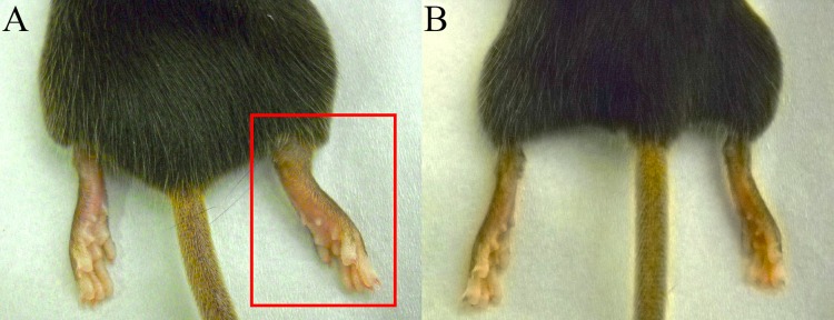
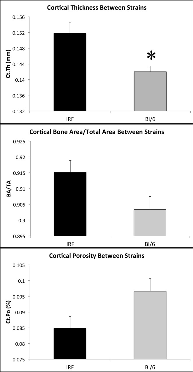
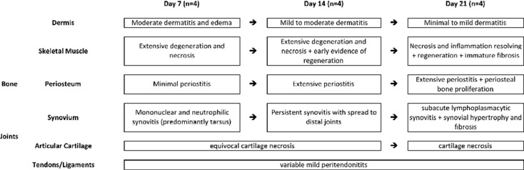
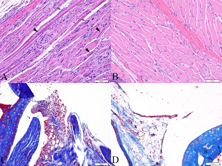
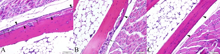
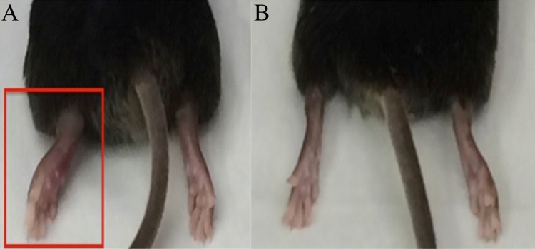
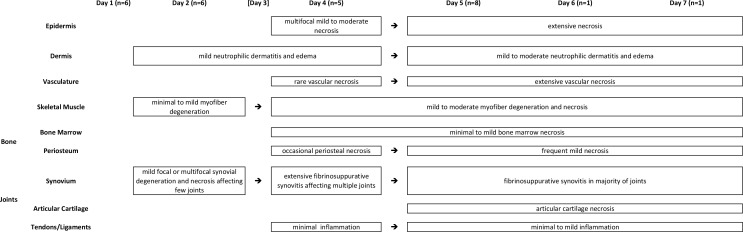
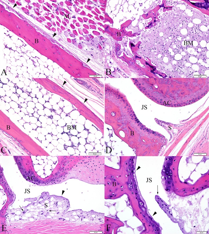
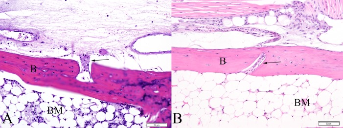
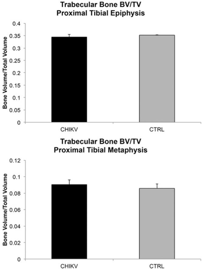
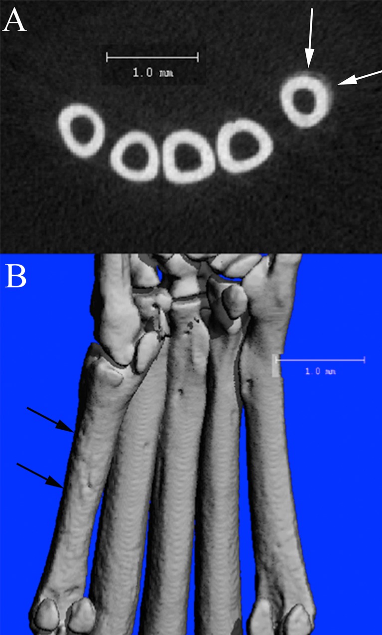
Chikungunya virus is an arbovirus spread predominantly by Aedes aegypti and Ae. albopictus mosquitoes, and causes debilitating arthralgia and arthritis. While these are common manifestations during acute infection and it has been suggested they can recur in patients chronically, gaps in knowledge regarding the pathogenesis still exist. Two established mouse models were utilized (adult IRF 3/7 -/- -/- and wild-type C57BL/6J mice) to evaluate disease manifestations in bones and joints at various timepoints. Novel lesions in C57BL/6J mice consisted of periostitis (91%) and foci of cartilage of necrosis (50% of mice at 21 DPI). Additionally, at 21 DPI, 50% and 75% of mice exhibited periosteal bone proliferation affecting the metatarsal bones, apparent via histology and μCT, respectively. μCT analysis did not reveal any alterations in trabecular bone volume measurements in C57BL/6J mice. Novel lesions demonstrated in IRF 3/7 -/- -/- mice at 5 DPI included focal regions of cartilage necrosis (20%), periosteal necrosis (66%), and multifocal ischemic bone marrow necrosis (100%). Contralateral feet in 100% of mice of both strains had similar, though milder lesions. Additionally, comparison of control IRF 3/7 -/- -/- and wild-type C57BL/6J mice demonstrated differences in cortical bone. These experiments demonstrate novel manifestations of disease similar to those occurring in humans, adding insight into disease pathogenesis, and representing new potential targets for therapeutic interventions. Additionally, results demonstrate the utility of μCT in studies of bone and joint pathology and illustrate differences in bone dynamics between mouse strains.
基孔肯雅病毒是一种虫媒病毒,主要通过埃及伊蚊和白纹伊蚊传播,可引起使人虚弱的关节痛和关节炎。虽然这些是急性感染期间的常见表现,并且有人认为它们可能在慢性患者中复发,但关于发病机制的知识仍存在空白。我们利用了两种已建立的小鼠模型(成年IRF 3/7 -/- -/- 和野生型C57BL/6J小鼠)来评估不同时间点骨骼和关节的疾病表现。C57BL/6J小鼠的新病变包括骨膜炎(91%)和软骨坏死灶(21天病程时50%的小鼠出现)。此外,在21天病程时,分别通过组织学和微计算机断层扫描(μCT)显示,50%和75%的小鼠出现影响跖骨的骨膜骨增生。μCT分析未发现C57BL/6J小鼠小梁骨体积测量有任何改变。在5天病程时,IRF 3/7 -/- -/- 小鼠出现的新病变包括局灶性软骨坏死区(20%)、骨膜坏死(66%)和多灶性缺血性骨髓坏死(100%)。两种品系100%的小鼠对侧足部有相似但较轻的病变。此外,对照IRF 3/7 -/- -/- 小鼠和野生型C57BL/6J小鼠的比较显示皮质骨存在差异。这些实验证明了与人类相似的疾病新表现,有助于深入了解疾病发病机制,并代表了治疗干预的新潜在靶点。此外,结果证明了μCT在骨骼和关节病理学研究中的实用性,并说明了小鼠品系之间骨动力学的差异。