Wang Yongming, Zou Zhiling, Song Hongwen, Xu Xiaodan, Wang Huijun, d'Oleire Uquillas Federico, Huang Xiting
School of Psychology, Southwest University Chongqing, China.
Department of Neurology, Massachusetts General Hospital, Harvard Medical School Boston, MA, USA.
Front Psychol. 2016 May 4;7:597. doi: 10.3389/fpsyg.2016.00597. eCollection 2016.
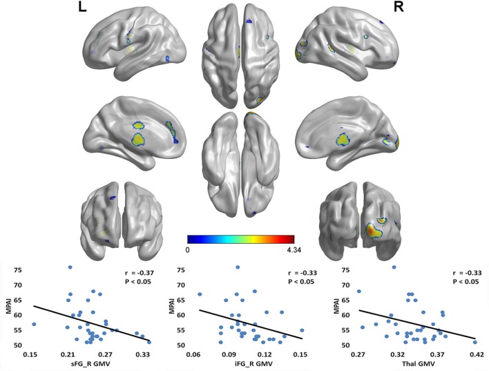
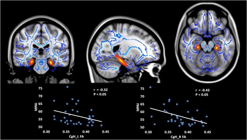
Mobile phone dependence (MPD) is a behavioral addiction that has become an increasing public mental health issue. While previous research has explored some of the factors that may predict MPD, the underlying neural mechanisms of MPD have not been investigated yet. The current study aimed to explore the microstructural variations associated with MPD as measured with functional Magnetic Resonance Imaging (fMRI). Gray matter volume (GMV) and white matter (WM) integrity [four indices: fractional anisotropy (FA); mean diffusivity (MD); axial diffusivity (AD); and radial diffusivity (RD)] were calculated via voxel-based morphometry (VBM) and tract-based spatial statistics (TBSS) analysis, respectively. Sixty-eight college students (42 female) were enrolled and separated into two groups [MPD group, N = 34; control group (CG), N = 34] based on Mobile Phone Addiction Index (MPAI) scale score. Trait impulsivity was also measured using the Barratt Impulsiveness Scale (BIS-11). In light of underlying trait impulsivity, results revealed decreased GMV in the MPD group relative to controls in regions such as the right superior frontal gyrus (sFG), right inferior frontal gyrus (iFG), and bilateral thalamus (Thal). In the MPD group, GMV in the above mentioned regions was negatively correlated with scores on the MPAI. Results also showed significantly less FA and AD measures of WM integrity in the MPD group relative to controls in bilateral hippocampal cingulum bundle fibers (CgH). Additionally, in the MPD group, FA of the CgH was also negatively correlated with scores on the MPAI. These findings provide the first morphological evidence of altered brain structure with mobile phone overuse, and may help to better understand the neural mechanisms of MPD in relation to other behavioral and substance addiction disorders.
手机依赖(MPD)是一种行为成瘾,已成为日益严重的公共心理健康问题。虽然先前的研究探讨了一些可能预测MPD的因素,但MPD潜在的神经机制尚未得到研究。当前的研究旨在探索与MPD相关的微观结构变化,通过功能磁共振成像(fMRI)进行测量。分别通过基于体素的形态计量学(VBM)和基于束的空间统计学(TBSS)分析计算灰质体积(GMV)和白质(WM)完整性[四个指标:分数各向异性(FA);平均扩散率(MD);轴向扩散率(AD);和径向扩散率(RD)]。招募了68名大学生(42名女性),并根据手机成瘾指数(MPAI)量表得分分为两组[MPD组,N = 34;对照组(CG),N = 34]。还使用巴拉特冲动量表(BIS-11)测量特质冲动性。鉴于潜在的特质冲动性,结果显示,相对于对照组,MPD组在右侧额上回(sFG)、右侧额下回(iFG)和双侧丘脑(Thal)等区域的GMV降低。在MPD组中,上述区域的GMV与MPAI得分呈负相关。结果还显示,相对于对照组,MPD组在双侧海马扣带束纤维(CgH)中的WM完整性的FA和AD测量值显著降低。此外,在MPD组中,CgH的FA也与MPAI得分呈负相关。这些发现提供了手机过度使用导致脑结构改变的首个形态学证据,并可能有助于更好地理解MPD与其他行为和物质成瘾障碍相关的神经机制。