Jia Xiaoqin, Li Xiaomin, Shen Yating, Miao Junjun, Liu Hao, Li Guoli, Wang Zhengbing
Department of Pathology, Medical College of Yangzhou University, Yangzhou, Jiangsu, China.
Department of Gastrointestinal Surgery, Clinical Medical College of Yangzhou University, Yangzhou, Jiangsu, China.
J Cell Mol Med. 2016 Oct;20(10):1898-907. doi: 10.1111/jcmm.12882. Epub 2016 May 31.
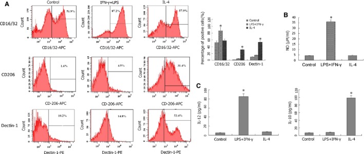
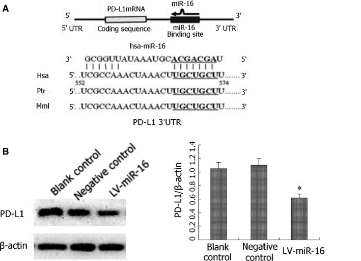
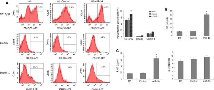
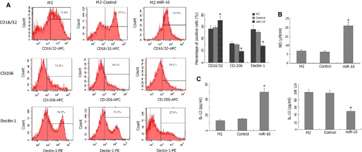
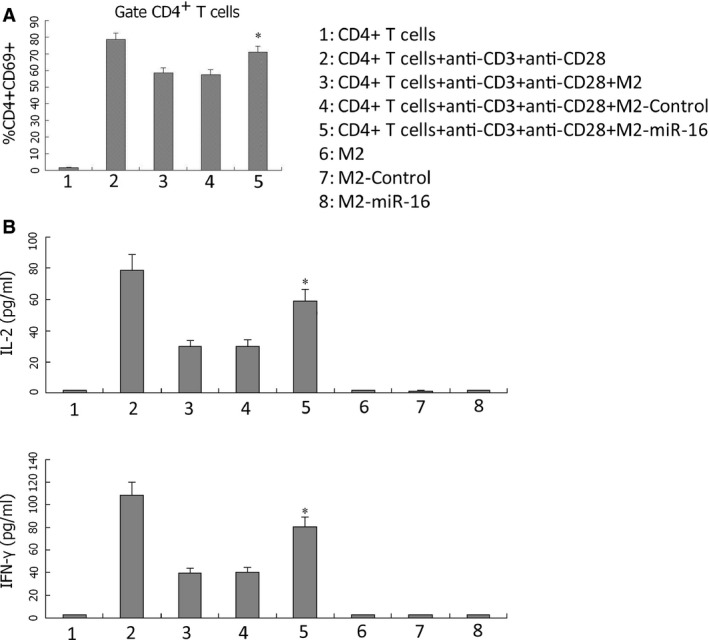
MiR-16 is a tumour suppressor that is down-regulated in certain human cancers. However, little is known on its activity in other cell types. In this study, we examined the biological significance and underlying mechanisms of miR-16 on macrophage polarization and subsequent T-cell activation. Mouse peritoneal macrophages were isolated and induced to undergo either M1 polarization with 100 ng/ml of interferon-γ and 20 ng/ml of lipopolysaccharide, or M2 polarization with 20 ng/ml of interleukin (IL)-4. The identity of polarized macrophages was determined by profiling cell-surface markers by flow cytometry and cytokine production by ELISA. Macrophages were infected with lentivirus-expressing miR-16 to assess the effects of miR-16. Effects on macrophage-T cell interactions were analysed by co-culturing purified CD4(+) T cells with miR-16-expressing peritoneal macrophages, and measuring activation marker CD69 by flow cytometry and cytokine secretion by ELISA. Bioinformatics analysis was applied to search for potential miR-16 targets and understand its underlying mechanisms. MiR-16-induced M1 differentiation of mouse peritoneal macrophages from either the basal M0- or M2-polarized state is indicated by the significant up-regulation of M1 marker CD16/32, repression of M2 marker CD206 and Dectin-1, and increased secretion of M1 cytokine IL-12 and nitric oxide. Consistently, miR-16-expressing macrophages stimulate the activation of purified CD4(+) T cells. Mechanistically, miR-16 significantly down-regulates the expression of PD-L1, a critical immune suppressor that controls macrophage-T cell interaction and T-cell activation. MiR-16 plays an important role in shifting macrophage polarization from M2 to M1 status, and functionally activating CD4(+) T cells. This effect is potentially mediated through the down-regulation of immune suppressor PD-L1.
微小RNA-16(miR-16)是一种肿瘤抑制因子,在某些人类癌症中表达下调。然而,其在其他细胞类型中的活性鲜为人知。在本研究中,我们探讨了miR-16对巨噬细胞极化及后续T细胞活化的生物学意义和潜在机制。分离小鼠腹腔巨噬细胞,用100 ng/ml干扰素-γ和20 ng/ml脂多糖诱导其向M1极化,或用20 ng/ml白细胞介素(IL)-4诱导其向M2极化。通过流式细胞术分析细胞表面标志物及ELISA检测细胞因子分泌来确定极化巨噬细胞的类型。用表达miR-16的慢病毒感染巨噬细胞以评估miR-16的作用。通过将纯化的CD4(+) T细胞与表达miR-16的腹腔巨噬细胞共培养,并用流式细胞术检测活化标志物CD69以及ELISA检测细胞因子分泌,来分析对巨噬细胞-T细胞相互作用的影响。应用生物信息学分析寻找潜在的miR-16靶标并了解其潜在机制。miR-16可诱导小鼠腹腔巨噬细胞从基础M0或M2极化状态向M1分化,表现为M1标志物CD16/32显著上调、M