• 文献检索
  • 文档翻译
  • 深度研究
  • 学术资讯
  • Suppr Zotero 插件Zotero 插件
  • 邀请有礼
  • 套餐&价格
  • 历史记录
应用&插件
Suppr Zotero 插件Zotero 插件浏览器插件Mac 客户端Windows 客户端微信小程序
定价
高级版会员购买积分包购买API积分包
服务
文献检索文档翻译深度研究API 文档MCP 服务
关于我们
关于 Suppr公司介绍联系我们用户协议隐私条款
关注我们

Suppr 超能文献

核心技术专利:CN118964589B侵权必究
粤ICP备2023148730 号-1Suppr @ 2026

文献检索

告别复杂PubMed语法,用中文像聊天一样搜索,搜遍4000万医学文献。AI智能推荐,让科研检索更轻松。

立即免费搜索

文件翻译

保留排版,准确专业,支持PDF/Word/PPT等文件格式,支持 12+语言互译。

免费翻译文档

深度研究

AI帮你快速写综述,25分钟生成高质量综述,智能提取关键信息,辅助科研写作。

立即免费体验

成年斑马鱼癌症进展与血管发育的定量体内光学断层扫描

Quantitative in vivo optical tomography of cancer progression & vasculature development in adult zebrafish.

作者信息

Kumar Sunil, Lockwood Nicola, Ramel Marie-Christine, Correia Teresa, Ellis Matthew, Alexandrov Yuriy, Andrews Natalie, Patel Rachel, Bugeon Laurence, Dallman Margaret J, Brandner Sebastian, Arridge Simon, Katan Matilda, McGinty James, Frankel Paul, French Paul M W

机构信息

Department of Physics, Imperial College London, London SW7 2AZ, UK.

Division of Medicine, University College London, London WC1E 6JF, UK.

出版信息

Oncotarget. 2016 Jul 12;7(28):43939-43948. doi: 10.18632/oncotarget.9756.

DOI:10.18632/oncotarget.9756
PMID:27259259
原文链接:https://pmc.ncbi.nlm.nih.gov/articles/PMC5190069/
Abstract

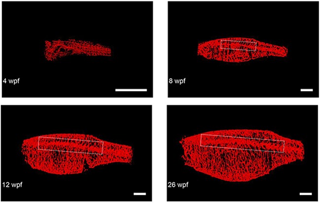
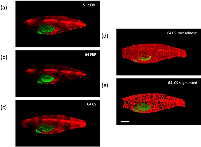
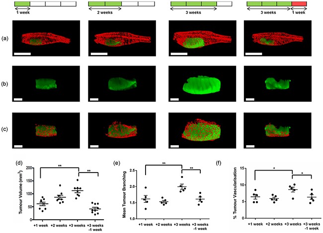
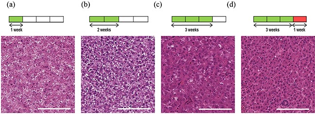
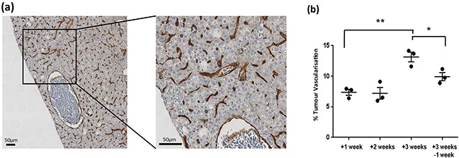
We describe a novel approach to study tumour progression and vasculature development in vivo via global 3-D fluorescence imaging of live non-pigmented adult zebrafish utilising angularly multiplexed optical projection tomography with compressive sensing (CS-OPT). This "mesoscopic" imaging method bridges a gap between established ~μm resolution 3-D fluorescence microscopy techniques and ~mm-resolved whole body planar imaging and diffuse tomography. Implementing angular multiplexing with CS-OPT, we demonstrate the in vivo global imaging of an inducible fluorescently labelled genetic model of liver cancer in adult non-pigmented zebrafish that also present fluorescently labelled vasculature. In this disease model, addition of a chemical inducer (doxycycline) drives expression of eGFP tagged oncogenic K-RASV12 in the liver of immune competent animals. We show that our novel in vivo global imaging methodology enables non-invasive quantitative imaging of the development of tumour and vasculature throughout the progression of the disease, which we have validated against established methods of pathology including immunohistochemistry. We have also demonstrated its potential for longitudinal imaging through a study of vascular development in the same zebrafish from early embryo to adulthood. We believe that this instrument, together with its associated analysis and data management tools, constitute a new platform for in vivo cancer studies and drug discovery in zebrafish disease models.

摘要

我们描述了一种通过利用具有压缩传感的角度复用光学投影断层扫描(CS-OPT)对活体非色素成年斑马鱼进行整体三维荧光成像来研究体内肿瘤进展和血管系统发育的新方法。这种“介观”成像方法弥合了既定的约微米分辨率的三维荧光显微镜技术与约毫米分辨率的全身平面成像和扩散断层扫描之间的差距。通过CS-OPT实施角度复用,我们展示了在成年非色素斑马鱼中对一种可诱导的荧光标记肝癌基因模型进行体内整体成像,该模型还呈现荧光标记的血管系统。在这个疾病模型中,添加化学诱导剂(强力霉素)可驱动免疫健全动物肝脏中eGFP标记的致癌K-RASV12的表达。我们表明,我们新颖的体内整体成像方法能够在疾病进展过程中对肿瘤和血管系统的发育进行非侵入性定量成像,我们已通过包括免疫组织化学在内的既定病理学方法对其进行了验证。我们还通过对同一斑马鱼从早期胚胎到成年期的血管发育研究证明了其纵向成像的潜力。我们相信,该仪器及其相关的分析和数据管理工具构成了一个用于斑马鱼疾病模型体内癌症研究和药物发现的新平台。

https://cdn.ncbi.nlm.nih.gov/pmc/blobs/0823/5190069/fd5cf6ff4f7b/oncotarget-07-43939-g006.jpg
https://cdn.ncbi.nlm.nih.gov/pmc/blobs/0823/5190069/d3282d9cfaa8/oncotarget-07-43939-g001.jpg
https://cdn.ncbi.nlm.nih.gov/pmc/blobs/0823/5190069/bc051700644e/oncotarget-07-43939-g002.jpg
https://cdn.ncbi.nlm.nih.gov/pmc/blobs/0823/5190069/3998481c7204/oncotarget-07-43939-g003.jpg
https://cdn.ncbi.nlm.nih.gov/pmc/blobs/0823/5190069/b98fe227cbc6/oncotarget-07-43939-g004.jpg
https://cdn.ncbi.nlm.nih.gov/pmc/blobs/0823/5190069/461b0f889803/oncotarget-07-43939-g005.jpg
https://cdn.ncbi.nlm.nih.gov/pmc/blobs/0823/5190069/fd5cf6ff4f7b/oncotarget-07-43939-g006.jpg

相似文献

1
Quantitative in vivo optical tomography of cancer progression & vasculature development in adult zebrafish.成年斑马鱼癌症进展与血管发育的定量体内光学断层扫描
Oncotarget. 2016 Jul 12;7(28):43939-43948. doi: 10.18632/oncotarget.9756.
2
Accelerated Optical Projection Tomography Applied to In Vivo Imaging of Zebrafish.加速光学投影断层扫描技术在斑马鱼活体成像中的应用
PLoS One. 2015 Aug 26;10(8):e0136213. doi: 10.1371/journal.pone.0136213. eCollection 2015.
3
In vivo label-free three-dimensional imaging of zebrafish vasculature with optical projection tomography.利用光学投影断层成像术对斑马鱼血管进行体内无标记的三维成像。
J Biomed Opt. 2011 Oct;16(10):100502. doi: 10.1117/1.3640808.
4
Single-shot optical projection tomography for high-speed volumetric imaging of dynamic biological samples.用于动态生物样本高速体积成像的单次光学投影断层扫描技术。
J Biophotonics. 2023 Feb;16(2):e202200232. doi: 10.1002/jbio.202200232. Epub 2022 Sep 29.
5
Comparison of optical projection tomography and light-sheet fluorescence microscopy.光学投影断层成像术与光片荧光显微镜的比较。
J Microsc. 2019 Jul;275(1):3-10. doi: 10.1111/jmi.12796. Epub 2019 Apr 30.
6
Quantitative measurement of blood velocity in zebrafish with optical vector field tomography.利用光学矢量场断层扫描技术对斑马鱼血液流速进行定量测量。
J Biophotonics. 2015 Jan;8(1-2):52-9. doi: 10.1002/jbio.201300162. Epub 2013 Dec 11.
7
Label-free optical projection tomography for quantitative three-dimensional anatomy of mouse embryo.无标记光学投影断层成像术用于定量分析小鼠胚胎的三维解剖结构
J Biophotonics. 2019 Jul;12(7):e201800481. doi: 10.1002/jbio.201800481. Epub 2019 Apr 29.
8
3D Optical Molecular Imaging of the Rodent Pancreas by OPT and LSFM.通过 OPT 和 LSFM 对啮齿动物胰腺进行 3D 光学分子成像。
Methods Mol Biol. 2023;2592:1-19. doi: 10.1007/978-1-0716-2807-2_1.
9
Time-gated optical projection tomography allows visualization of adult zebrafish internal structures.时门控光学投影断层成像术可用于可视化成年斑马鱼的内部结构。
PLoS One. 2012;7(11):e50744. doi: 10.1371/journal.pone.0050744. Epub 2012 Nov 19.
10
An inducible kras(V12) transgenic zebrafish model for liver tumorigenesis and chemical drug screening.诱导型 Kras(V12)转基因斑马鱼模型用于肝肿瘤发生和化学药物筛选。
Dis Model Mech. 2012 Jan;5(1):63-72. doi: 10.1242/dmm.008367. Epub 2011 Sep 8.

引用本文的文献

1
Optical projection tomography implemented for accessibility and low cost ().为实现可及性和低成本而实施的光学投影断层扫描()。 (括号部分原文缺失具体内容)
Philos Trans A Math Phys Eng Sci. 2024 Jul 9;382(2274):20230101. doi: 10.1098/rsta.2023.0101. Epub 2024 Jun 3.
2
Smad4 controls signaling robustness and morphogenesis by differentially contributing to the Nodal and BMP pathways.Smad4 通过对 Nodal 和 BMP 途径的不同贡献来控制信号稳健性和形态发生。
Nat Commun. 2021 Nov 4;12(1):6374. doi: 10.1038/s41467-021-26486-3.
3
Cerebrovascular development: mechanisms and experimental approaches.

本文引用的文献

1
Accelerated Optical Projection Tomography Applied to In Vivo Imaging of Zebrafish.加速光学投影断层扫描技术在斑马鱼活体成像中的应用
PLoS One. 2015 Aug 26;10(8):e0136213. doi: 10.1371/journal.pone.0136213. eCollection 2015.
2
A Quantitative System for Studying Metastasis Using Transparent Zebrafish.一种使用透明斑马鱼研究转移的定量系统。
Cancer Res. 2015 Oct 15;75(20):4272-4282. doi: 10.1158/0008-5472.CAN-14-3319. Epub 2015 Aug 17.
3
Zebrafish: a new companion for translational research in oncology.斑马鱼:肿瘤转化研究的新伙伴。
脑血管发育:机制与实验方法。
Cell Mol Life Sci. 2021 May;78(9):4377-4398. doi: 10.1007/s00018-021-03790-1. Epub 2021 Mar 10.
4
P130Cas/bcar1 mediates zebrafish caudal vein plexus angiogenesis.P130Cas/bcar1 介导斑马鱼尾静脉丛血管生成。
Sci Rep. 2020 Sep 24;10(1):15589. doi: 10.1038/s41598-020-71753-w.
5
OptiJ: Open-source optical projection tomography of large organ samples.OptiJ:开源大型器官样本光学投影层析成像。
Sci Rep. 2019 Oct 30;9(1):15693. doi: 10.1038/s41598-019-52065-0.
6
Gaussian Light Model in Brightfield Optical Projection Tomography.明场光学投影层析中的高斯光模型。
Sci Rep. 2019 Sep 26;9(1):13934. doi: 10.1038/s41598-019-50469-6.
7
Convolutional neural networks for reconstruction of undersampled optical projection tomography data applied to in vivo imaging of zebrafish.应用于斑马鱼体内成像的欠采样光学投影断层成像数据重建的卷积神经网络。
J Biophotonics. 2019 Dec;12(12):e201900128. doi: 10.1002/jbio.201900128. Epub 2019 Aug 29.
8
Slice-illuminated optical projection tomography.切片照明光学投影层析成像。
Opt Lett. 2018 Nov 15;43(22):5555-5558. doi: 10.1364/OL.43.005555.
9
Optimal Anesthetic Regime for Motionless Three-Dimensional Image Acquisition During Longitudinal Studies of Adult Nonpigmented Zebrafish.成年非色素斑马鱼纵向研究中用于静止三维图像采集的最佳麻醉方案
Zebrafish. 2017 Apr;14(2):133-139. doi: 10.1089/zeb.2016.1343. Epub 2017 Jan 30.
Clin Cancer Res. 2015 Mar 1;21(5):969-75. doi: 10.1158/1078-0432.CCR-14-2921. Epub 2015 Jan 8.
4
Xmrk, kras and myc transgenic zebrafish liver cancer models share molecular signatures with subsets of human hepatocellular carcinoma.Xmrk、kras和myc转基因斑马鱼肝癌模型与人类肝细胞癌亚群具有共同的分子特征。
PLoS One. 2014 Mar 14;9(3):e91179. doi: 10.1371/journal.pone.0091179. eCollection 2014.
5
Crosstalk of Ras and Rho: activation of RhoA abates Kras-induced liver tumorigenesis in transgenic zebrafish models.Ras 和 Rho 之间的串扰:RhoA 的激活减轻了转基因斑马鱼模型中 Kras 诱导的肝肿瘤发生。
Oncogene. 2014 May 22;33(21):2717-27. doi: 10.1038/onc.2013.240. Epub 2013 Jul 1.
6
Simultaneous angular multiplexing optical projection tomography at shifted focal planes.在移焦平面上进行同时角度复用光学投影层析成像。
Opt Lett. 2013 Mar 15;38(6):851-3. doi: 10.1364/OL.38.000851.
7
Non-invasive whole-body imaging of adult zebrafish with optoacoustic tomography.采用光声断层成像技术对成年斑马鱼进行无创性全身成像。
Phys Med Biol. 2012 Nov 21;57(22):7227-37. doi: 10.1088/0031-9155/57/22/7227. Epub 2012 Oct 17.
8
OMERO: flexible, model-driven data management for experimental biology.OMERO:用于实验生物学的灵活、模型驱动的数据管理。
Nat Methods. 2012 Feb 28;9(3):245-53. doi: 10.1038/nmeth.1896.
9
An inducible kras(V12) transgenic zebrafish model for liver tumorigenesis and chemical drug screening.诱导型 Kras(V12)转基因斑马鱼模型用于肝肿瘤发生和化学药物筛选。
Dis Model Mech. 2012 Jan;5(1):63-72. doi: 10.1242/dmm.008367. Epub 2011 Sep 8.
10
In vivo fluorescence lifetime optical projection tomography.体内荧光寿命光学投影断层成像。
Biomed Opt Express. 2011 Apr 26;2(5):1340-50. doi: 10.1364/BOE.2.001340.