Thomsen Barbara, Garosi Laurent, Skerritt Geoff, Rusbridge Clare, Sparrow Tim, Berendt Mette, Gredal Hanne
Department of Veterinary Clinical and Animal Sciences, University Hospital for Companion Animals, University of Copenhagen, Dyrlægevej 16, 1870, Frederiksberg C, Denmark.
Davies Veterinary Specialists, Manor Farm Business Park, Higham Gobion, Hitchin, England, SG5 3HR, UK.
Acta Vet Scand. 2016 Jun 7;58(1):40. doi: 10.1186/s13028-016-0219-2.
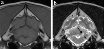
In dogs with ischaemic stroke, a very common site of infarction is the cerebellum. The aim of this study was to characterise neurological signs in relation to infarct topography in dogs with suspected cerebellar ischaemic stroke and to report short-term outcome confined to the hospitalisation period. A retrospective multicentre study of dogs with suspected cerebellar ischaemic stroke examined from 2010-2015 at five veterinary referral hospitals was performed. Findings from clinical, neurological, and paraclinical investigations including magnetic resonance imaging were assessed.
Twenty-three dogs, 13 females and 10 males with a median age of 8 years and 8 months, were included in the study. The Cavalier King Charles Spaniel (n = 9) was a commonly represented breed. All ischaemic strokes were located to the vascular territory of the rostral cerebellar artery including four extensive and 19 limited occlusions. The most prominent neurological deficits were gait abnormalities (ataxia with hypermetria n = 11, ataxia without hypermetria n = 4, non-ambulatory n = 6), head tilt (n = 13), nystagmus (n = 8), decreased menace response (n = 7), postural reaction deficits (n = 7), and proprioceptive deficits (n = 5). Neurological signs appeared irrespective of the infarct being classified as extensive or limited. All dogs survived and were discharged within 1-10 days of hospitalisation.
Dogs affected by rostral cerebellar ischaemic stroke typically present with a collection of neurological deficits characterised by ataxia, head tilt, and nystagmus irrespective of the specific cerebellar infarct topography. In dogs with peracute to acute onset of these neurological deficits, cerebellar ischaemic stroke should be considered an important differential diagnosis, and neuroimaging investigations are indicated. Although dogs are often severely compromised at presentation, short-term prognosis is excellent and rapid clinical improvement may be observed within the first week following the ischaemic stroke.
在患有缺血性中风的犬类中,梗死的一个非常常见部位是小脑。本研究的目的是描述疑似小脑缺血性中风犬类的神经体征与梗死灶地形学的关系,并报告仅限于住院期间的短期预后情况。对2010年至2015年在五家兽医转诊医院检查的疑似小脑缺血性中风犬类进行了一项回顾性多中心研究。评估了包括磁共振成像在内的临床、神经和辅助临床检查结果。
23只犬类纳入研究,其中13只为雌性,10只为雄性,中位年龄为8岁8个月。骑士查理王小猎犬(n = 9)是常见品种。所有缺血性中风均位于小脑前动脉的血管区域,包括4例广泛闭塞和19例局限性闭塞。最突出的神经功能缺损为步态异常(共济失调伴辨距过度n = 11,共济失调不伴辨距过度n = 4,不能行走n = 6)、头部倾斜(n = 13)、眼球震颤(n = 8)、威胁反应减弱(n = 7)、姿势反应缺陷(n = 7)和本体感觉缺陷(n = 5)。无论梗死灶被分类为广泛型还是局限型,神经体征均会出现。所有犬类均存活并在住院1至10天内出院。
受小脑前缺血性中风影响的犬类通常表现出一系列以共济失调、头部倾斜和眼球震颤为特征的神经功能缺损,而与特定的小脑梗死灶地形学无关。在出现这些神经功能缺损的超急性至急性发作的犬类中,应将小脑缺血性中风视为重要的鉴别诊断,并建议进行神经影像学检查。尽管犬类在就诊时通常严重受损,但短期预后良好,在缺血性中风后的第一周内可能会观察到临床迅速改善。