Mendell Ari L, Atwi Sarah, Bailey Craig D C, McCloskey Dan, Scharfman Helen E, MacLusky Neil J
Department of Biomedical Sciences, Ontario Veterinary College, University of Guelph, Guelph, ON, N1G 2W1, Canada.
Nathan Kline Institute for Psychiatric Research, Center of Dementia Research, Orangeburg, NY, 10962, USA.
Brain Struct Funct. 2017 Jan;222(1):587-601. doi: 10.1007/s00429-016-1237-6. Epub 2016 Jun 9.
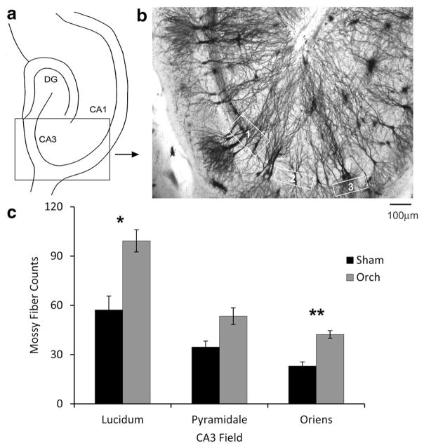
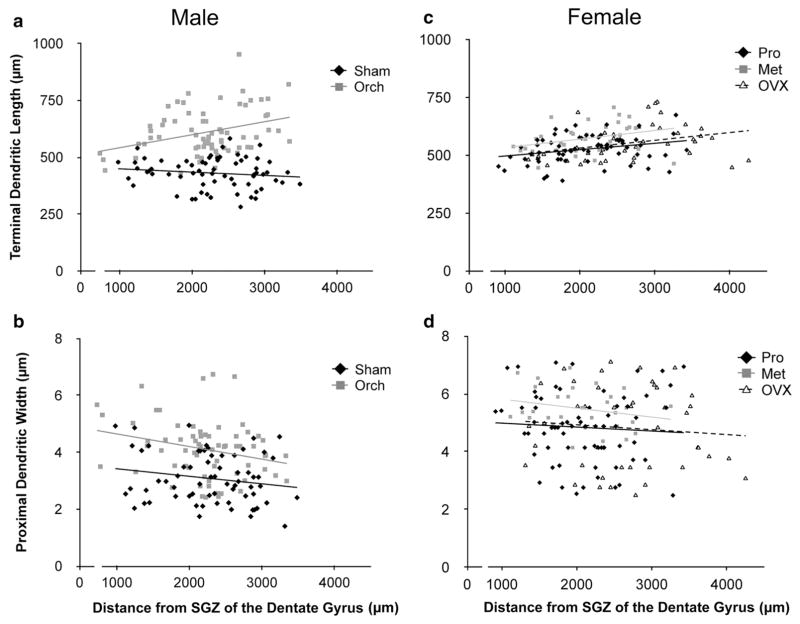
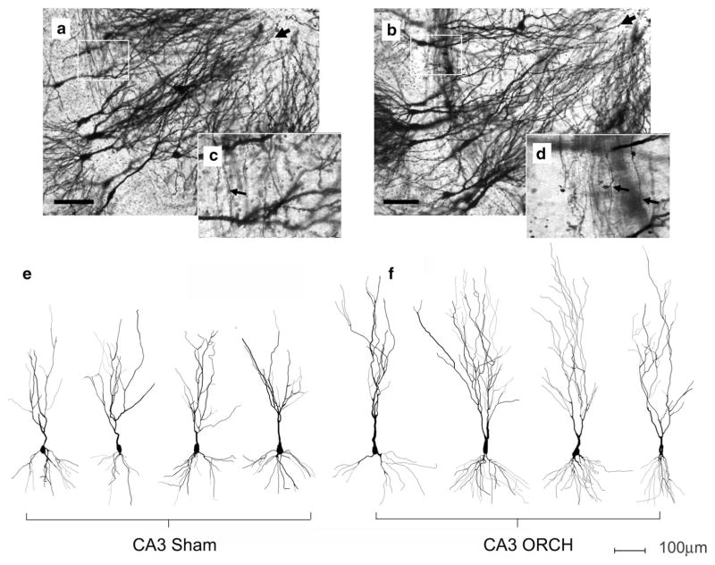
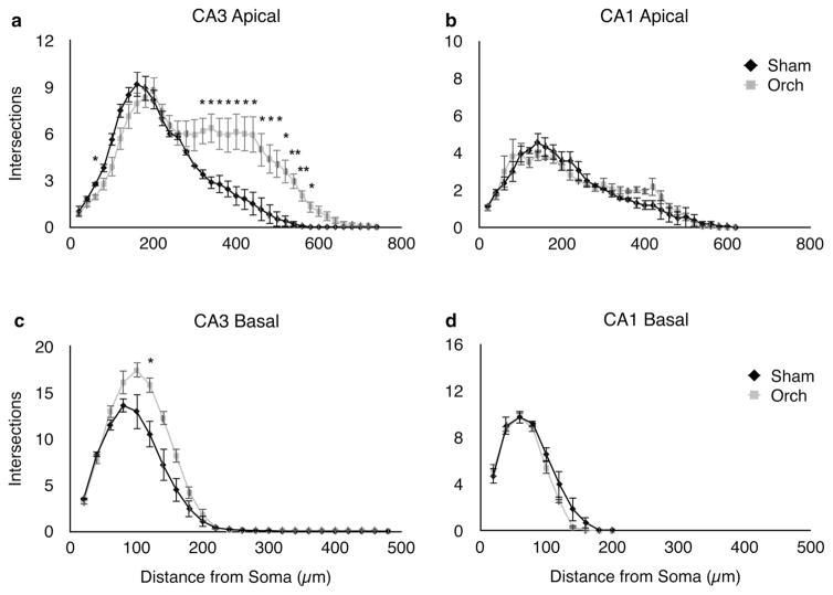
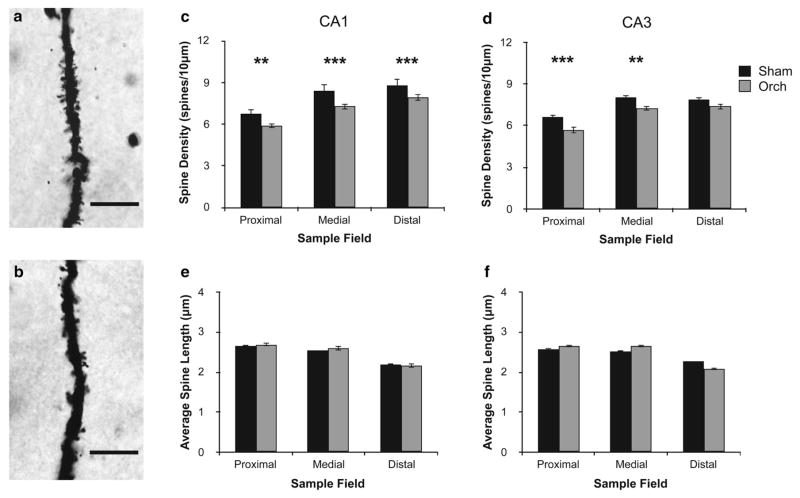
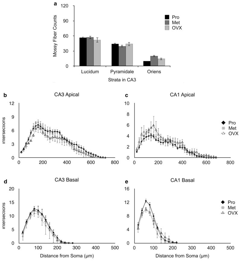
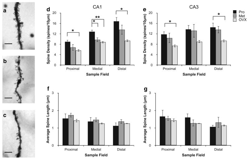
Androgen loss is an important clinical concern because of its cognitive and behavioral effects. Changes in androgen levels are also suspected to contribute to neurological disease. However, the available data on the effects of androgen deprivation in areas of the brain that are central to cognition, like the hippocampus, are mixed. In this study, morphological analysis of pyramidal cells was used to investigate if structural changes could potentially contribute to the mixed cognitive effects that have been observed after androgen loss in males. Male Sprague-Dawley rats were orchidectomized or sham-operated. Two months later, their brains were Golgi-impregnated for morphological analysis. Morphological endpoints were studied in areas CA3 and CA1, with comparisons to females either intact or 2 months after ovariectomy. CA3 pyramidal neurons of orchidectomized rats exhibited marked increases in apical dendritic arborization. There were increases in mossy fiber afferent density in area CA3, as well as robust enhancements to dendritic structure in area CA3 of orchidectomized males, but not in CA1. Remarkably, apical dendritic length of CA3 pyramidal cells increased, while spine density declined. By contrast, in females overall dendritic structure was minimally affected by ovariectomy, while dendritic spine density was greatly reduced. Sex differences and subfield-specific effects of gonadal hormone deprivation on the hippocampal circuitry may help explain the different behavioral effects reported in males and females after gonadectomy, or other conditions associated with declining gonadal hormone secretion.
雄激素丧失因其对认知和行为的影响而成为一个重要的临床关注点。雄激素水平的变化也被怀疑与神经疾病有关。然而,关于雄激素剥夺对大脑中对认知至关重要的区域(如海马体)影响的现有数据并不一致。在本研究中,使用锥体细胞的形态学分析来调查结构变化是否可能导致在雄性雄激素丧失后观察到的混合认知效应。将雄性Sprague-Dawley大鼠进行去势或假手术。两个月后,将它们的大脑进行高尔基染色以进行形态学分析。在CA3区和CA1区研究形态学终点,并与完整雌性或卵巢切除术后2个月的雌性进行比较。去势大鼠的CA3锥体细胞顶端树突分支显著增加。CA3区苔藓纤维传入密度增加,去势雄性大鼠CA3区的树突结构也有显著增强,但CA1区没有。值得注意的是,CA3锥体细胞的顶端树突长度增加,而棘突密度下降。相比之下,在雌性中,卵巢切除术对整体树突结构的影响最小,而树突棘密度则大大降低。性腺激素剥夺对海马回路的性别差异和亚区特异性影响可能有助于解释在性腺切除术后或其他与性腺激素分泌下降相关的情况下,雄性和雌性报告的不同行为效应。