Grawe Brian M, Williams Phillip N, Burge Alissa, Voigt Marcia, Altchek David W, Hannafin Jo A, Allen Answorth A
Sports Medicine and Shoulder Reconstruction, Department of Orthopaedics, University of Cincinnati Academic Health Center, Cincinnati, Ohio, USA.
Kerlan-Jobe Orthopaedic Clinic, Los Angeles, California, USA.
Orthop J Sports Med. 2016 May 26;4(5):2325967116646360. doi: 10.1177/2325967116646360. eCollection 2016 May.
Recent clinical investigations have identified inadequate autograft hamstring graft diameter (<8 mm) to be predictive of failure after reconstruction of the anterior cruciate ligament (ACL).
PURPOSE/HYPOTHESIS: The objective of this study was to determine the utility of preoperative magnetic resonance imaging (MRI) variables of the hamstring tendons for the prediction of graft diameter at the time of surgery. The hypothesis was that cross-sectional area (CSA) of the hamstring tendon measured on MRI could accurately predict graft diameter, and threshold measurements could be established to predict graft diameter at the time of surgery.
Cohort study (diagnosis); Level of evidence, 2.
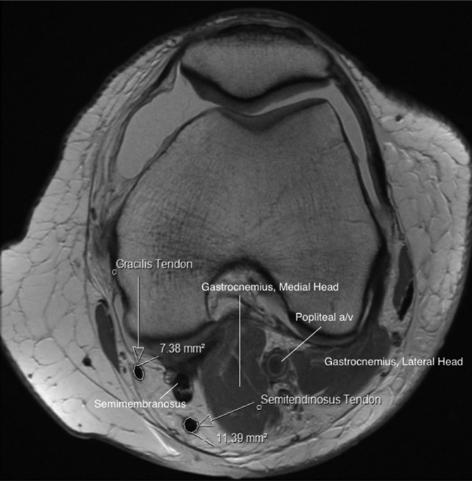
A total of 84 consecutive skeletally mature patients prospectively enrolled in our ACL reconstruction patient registry were identified for study purposes. Patients were included if they underwent an MRI of the affected knee at our institution prior to ACL reconstruction with hamstring (HT) autograft. Graft preparation was performed via a standard quadrupled hamstring technique after harvesting both the gracilis and semitendinosus (4-GST). The smallest diameter end of the HT autograft was then utilized for measurement analysis. Total CSA was calculated for both hamstring tendons using the "region of interest tool" on the corresponding proton density-weighted axial image of the knee at the widest condylar dimension. Three independent reviewers measured the MRI scans so that intra- and interrater reliability of the measurements could be determined. A trend analysis was then undertaken to establish correlations between the MRI CSA and graft diameter. Predictive analysis was then performed to establish threshold MRI measurement values for specific graft diameters and determine whether any patient-specific factors would affect graft diameter (age, sex, and body mass index).
Mean patient age at the time of surgery was 36 years (range, 11-57 years). Intra- and interrater reliability measurements achieved near-perfect agreement for CSA measurements, with intraclass correlation coefficients (ICCs) of 0.994 and 0.932, respectively. Trend analysis demonstrated that increasing CSA correlated well with increasing overall diameter of the graft (P < .001). Receiver operating characteristic (ROC) curves were generated to evaluate threshold CSA measurements for various graft diameters. Maximum sensitivity values of 21.64, 25.25, and 28.256 mm(2) were established for the respective graft diameters of 8, 9, and 10 mm in the 4-GST group. Independent patient factors of younger age, shorter stature, and female sex were significantly associated with graft diameter (P = .019, .034, and .028, respectively).
Preoperative MRI can be used to accurately predict quadrupled hamstring autograft diameter at the time of surgery. A total cross-sectional area of >22 mm(2) can reliably provide a graft diameter of >8 mm at the time of surgery.
近期临床研究发现,自体腘绳肌移植物直径不足(<8 mm)可预测前交叉韧带(ACL)重建术后失败。
目的/假设:本研究的目的是确定术前腘绳肌腱的磁共振成像(MRI)变量对手术时移植物直径的预测作用。假设是,MRI测量的腘绳肌腱横截面积(CSA)可准确预测移植物直径,并可建立阈值测量值以预测手术时的移植物直径。
队列研究(诊断);证据等级,2级。
共有84例连续的骨骼成熟患者前瞻性纳入我们的ACL重建患者登记系统,用于研究目的。如果患者在我们机构接受自体腘绳肌(HT)移植重建ACL之前对患膝进行了MRI检查,则纳入研究。在采集股薄肌和半腱肌(4-GST)后,通过标准的四股腘绳肌技术进行移植物制备。然后使用HT自体移植物的最小直径端进行测量分析。使用膝关节相应质子密度加权轴向图像上最宽髁尺寸处的“感兴趣区域工具”计算两条腘绳肌腱的总CSA。由三名独立的审阅者测量MRI扫描图像,以便确定测量的组内和组间可靠性。然后进行趋势分析,以建立MRI CSA与移植物直径之间的相关性。然后进行预测分析,以建立特定移植物直径的阈值MRI测量值,并确定是否有任何患者特定因素会影响移植物直径(年龄、性别和体重指数)。
手术时患者的平均年龄为36岁(范围11 - 57岁)。组内和组间可靠性测量在CSA测量方面达到了近乎完美的一致性,组内相关系数(ICC)分别为0.994和0.932。趋势分析表明,CSA增加与移植物总体直径增加密切相关(P <.001)。生成受试者操作特征(ROC)曲线以评估各种移植物直径的阈值CSA测量值。在4-GST组中,对于8、9和10 mm的移植物直径,分别建立了最大敏感性值21.64、25.25和28.256 mm²。年龄较小、身材较矮和女性等独立患者因素与移植物直径显著相关(分别为P =.019、.034和.028)。
术前MRI可用于准确预测手术时四股自体腘绳肌移植物的直径。总横截面积>22 mm²可在手术时可靠地提供直径>8 mm的移植物。