Guadiana Sarah M, Parker Alexander K, Filho Gileno F, Sequeira Ashton, Semple-Rowland Susan, Shaw Gerry, Mandel Ronald J, Foster Thomas C, Kumar Ashok, Sarkisian Matthew R
Department of Neuroscience, McKnight Brain Institute, University of Florida Gainesville, FL, USA.
Department of Neuroscience, McKnight Brain Institute, University of FloridaGainesville, FL, USA; EnCor Biotechnology Inc.Gainesville, FL, USA.
Front Aging Neurosci. 2016 May 31;8:127. doi: 10.3389/fnagi.2016.00127. eCollection 2016.
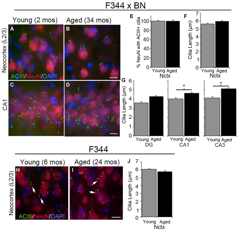
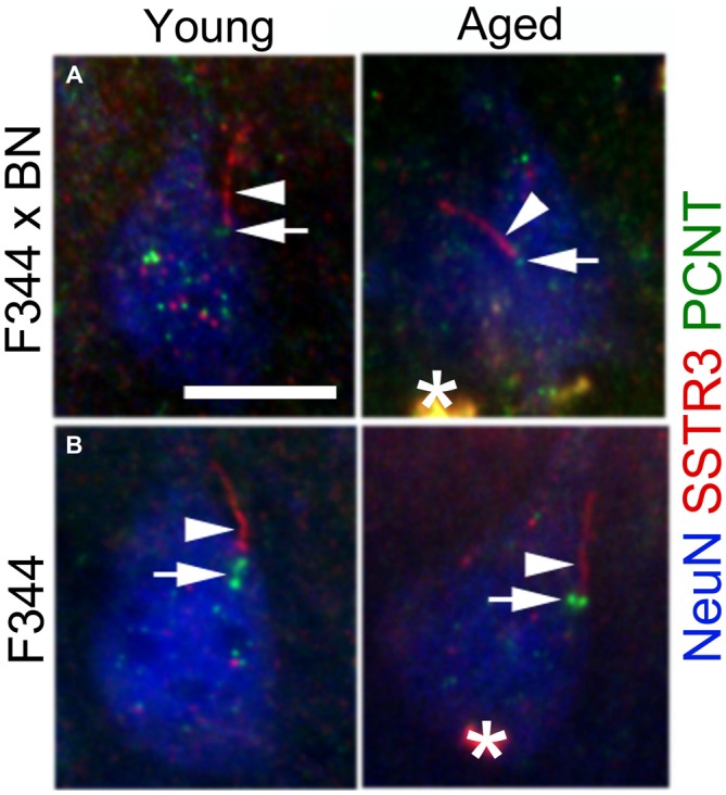
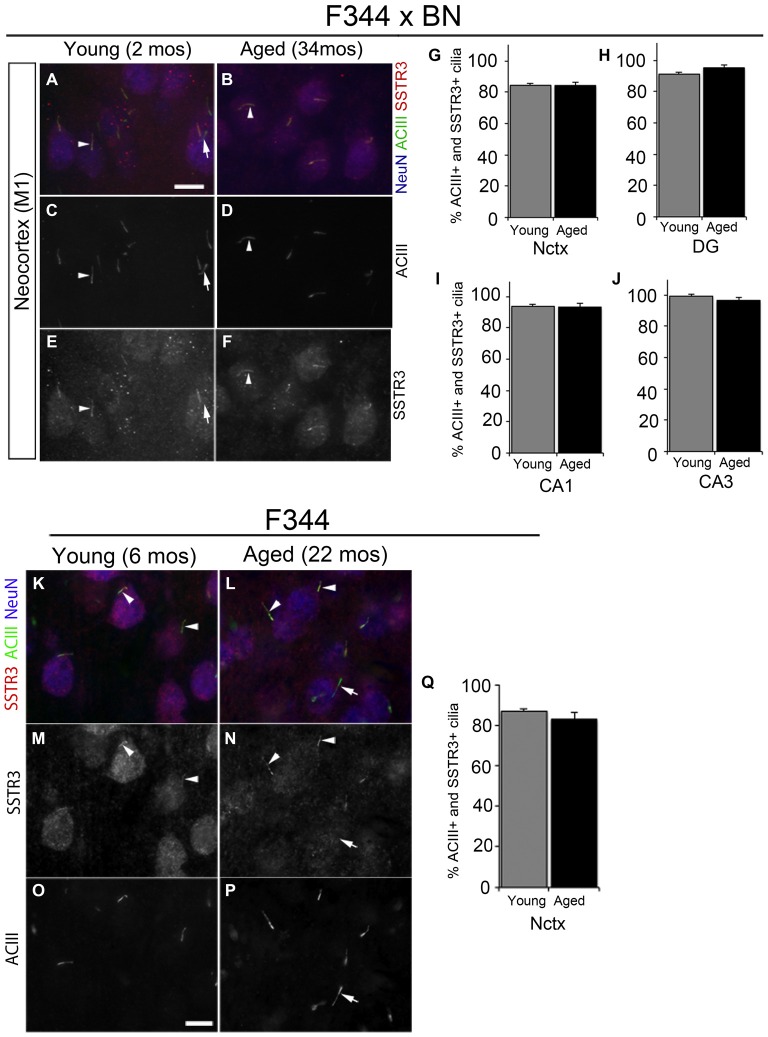
The primary cilia of forebrain neurons assemble around birth and become enriched with neuromodulatory receptors. Our understanding of the permanence of these structures and their associated signaling pathways in the aging brain is poor, but they are worthy of investigation because disruptions in neuronal cilia signaling have been implicated in changes in learning and memory, depression-like symptoms, and sleep anomalies. Here, we asked whether neurons in aged forebrain retain primary cilia and whether the staining characteristics of aged cilia for type 3 adenylyl cyclase (ACIII), somatostatin receptor 3 (SSTR3), and pericentrin resemble those of cilia in younger forebrain. To test this, we analyzed immunostained sections of forebrain tissues taken from young and aged male Fischer 344 (F344) and F344 × Brown Norway (F344 × BN) rats. Analyses of ACIII and SSTR3 in young and aged cortices of both strains of rats revealed that the staining patterns in the neocortex and hippocampus were comparable. Virtually every NeuN positive cell examined possessed an ACIII positive cilium. The lengths of ACIII positive cilia in neocortex were similar between young and aged for both strains, whereas in F344 × BN hippocampus, the cilia lengths increased with age in CA1 and CA3, but not in dentate gyrus (DG). Additionally, the percentages of ACIII positive cilia that were also SSTR3 positive did not differ between young and aged tissues in either strain. We also found that pericentrin, a protein that localizes to the basal bodies of neuronal cilia and functions in primary cilia assembly, persisted in aged cortical neurons of both rat strains. Collectively, our data show that neurons in aged rat forebrain possess primary cilia and that these cilia, like those present in younger brain, continue to localize ACIII, SSTR3, and pericentrin. Further studies will be required to determine if the function and signaling pathways regulated by cilia are similar in aged compared to young brain.
前脑神经元的初级纤毛在出生前后组装,并富含神经调节受体。我们对这些结构及其相关信号通路在衰老大脑中的持久性了解甚少,但它们值得研究,因为神经元纤毛信号传导的破坏与学习和记忆的变化、抑郁样症状以及睡眠异常有关。在这里,我们询问衰老前脑中的神经元是否保留初级纤毛,以及衰老纤毛对3型腺苷酸环化酶(ACIII)、生长抑素受体3(SSTR3)和中心体蛋白的染色特征是否与年轻前脑中的纤毛相似。为了测试这一点,我们分析了取自年轻和老年雄性Fischer 344(F344)和F344×Brown Norway(F344×BN)大鼠的前脑组织免疫染色切片。对两种品系大鼠年轻和老年皮质中ACIII和SSTR3的分析表明,新皮质和海马体中的染色模式具有可比性。几乎每个检测的NeuN阳性细胞都有一个ACIII阳性纤毛。两种品系的新皮质中ACIII阳性纤毛的长度在年轻和老年之间相似,而在F344×BN海马体中,CA1和CA3区的纤毛长度随年龄增加,齿状回(DG)中则不然。此外,两种品系的年轻和老年组织中ACIII阳性纤毛同时也是SSTR3阳性的百分比没有差异。我们还发现,中心体蛋白是一种定位于神经元纤毛基体并在初级纤毛组装中起作用的蛋白质,在两种大鼠品系的老年皮质神经元中持续存在。总的来说,我们的数据表明,老年大鼠前脑中的神经元拥有初级纤毛,并且这些纤毛与年轻大脑中的纤毛一样,继续定位ACIII、SSTR3和中心体蛋白。需要进一步研究以确定与年轻大脑相比,衰老大脑中纤毛调节的功能和信号通路是否相似。