• 文献检索
  • 文档翻译
  • 深度研究
  • 学术资讯
  • Suppr Zotero 插件Zotero 插件
  • 邀请有礼
  • 套餐&价格
  • 历史记录
应用&插件
Suppr Zotero 插件Zotero 插件浏览器插件Mac 客户端Windows 客户端微信小程序
定价
高级版会员购买积分包购买API积分包
服务
文献检索文档翻译深度研究API 文档MCP 服务
关于我们
关于 Suppr公司介绍联系我们用户协议隐私条款
关注我们

Suppr 超能文献

核心技术专利:CN118964589B侵权必究
粤ICP备2023148730 号-1Suppr @ 2026

文献检索

告别复杂PubMed语法,用中文像聊天一样搜索,搜遍4000万医学文献。AI智能推荐,让科研检索更轻松。

立即免费搜索

文件翻译

保留排版,准确专业,支持PDF/Word/PPT等文件格式,支持 12+语言互译。

免费翻译文档

深度研究

AI帮你快速写综述,25分钟生成高质量综述,智能提取关键信息,辅助科研写作。

立即免费体验

大鼠体内铜纳米颗粒和铜离子的比较毒性及生物分布

Comparative toxicity and biodistribution of copper nanoparticles and cupric ions in rats.

作者信息

Lee In-Chul, Ko Je-Won, Park Sung-Hyeuk, Lim Je-Oh, Shin In-Sik, Moon Changjong, Kim Sung-Hwan, Heo Jeong-Doo, Kim Jong-Choon

机构信息

College of Veterinary Medicine BK21 Plus Project Team, Chonnam National University, Gwangju, Republic of Korea.

Jeonbuk Department of Inhalation Research, Korea Institute of Toxicology, Jeongeup, Republic of Korea.

出版信息

Int J Nanomedicine. 2016 Jun 16;11:2883-900. doi: 10.2147/IJN.S106346. eCollection 2016.

DOI:10.2147/IJN.S106346
PMID:27366066
原文链接:https://pmc.ncbi.nlm.nih.gov/articles/PMC4913985/
Abstract

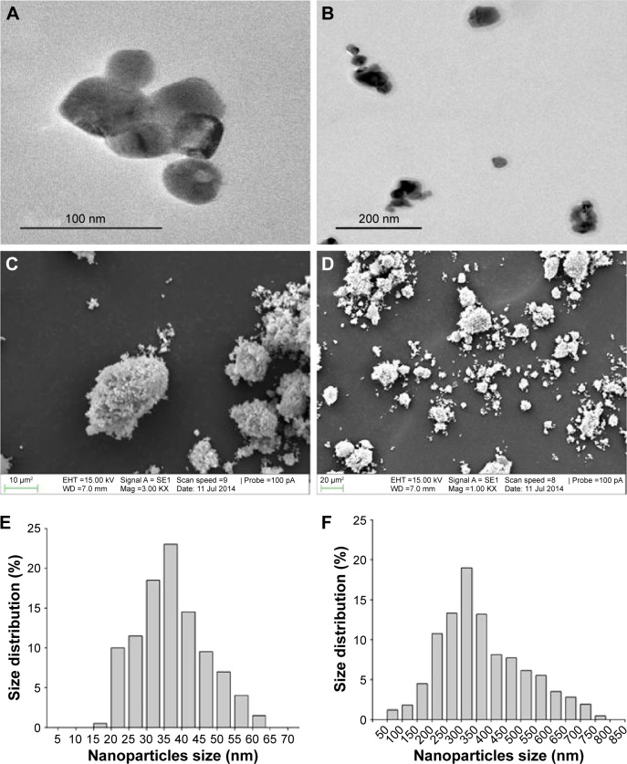
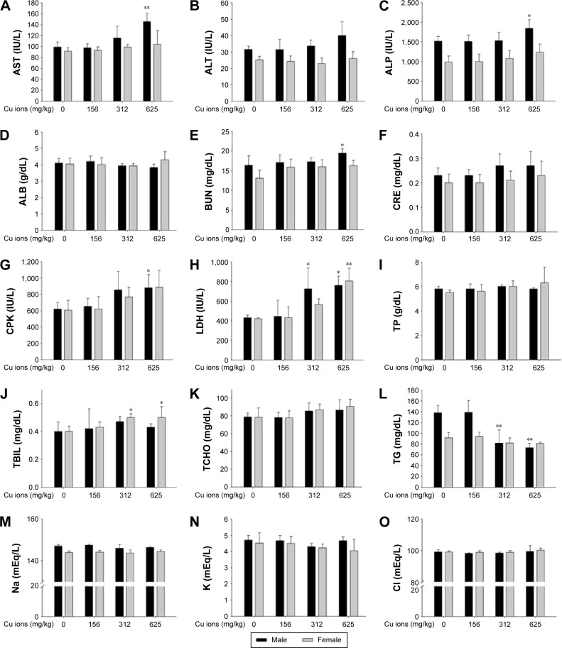
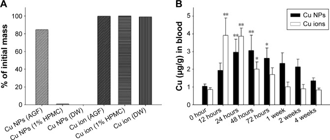
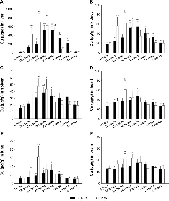
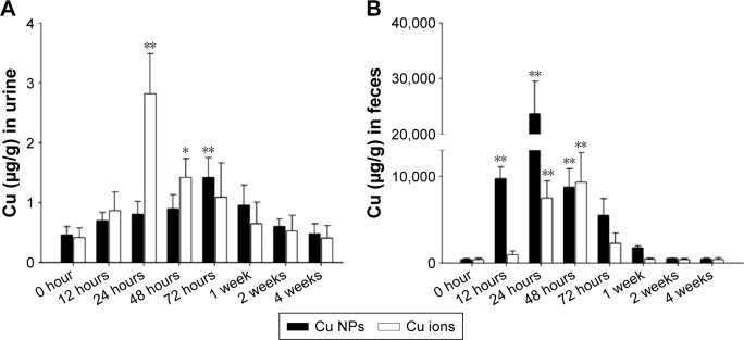
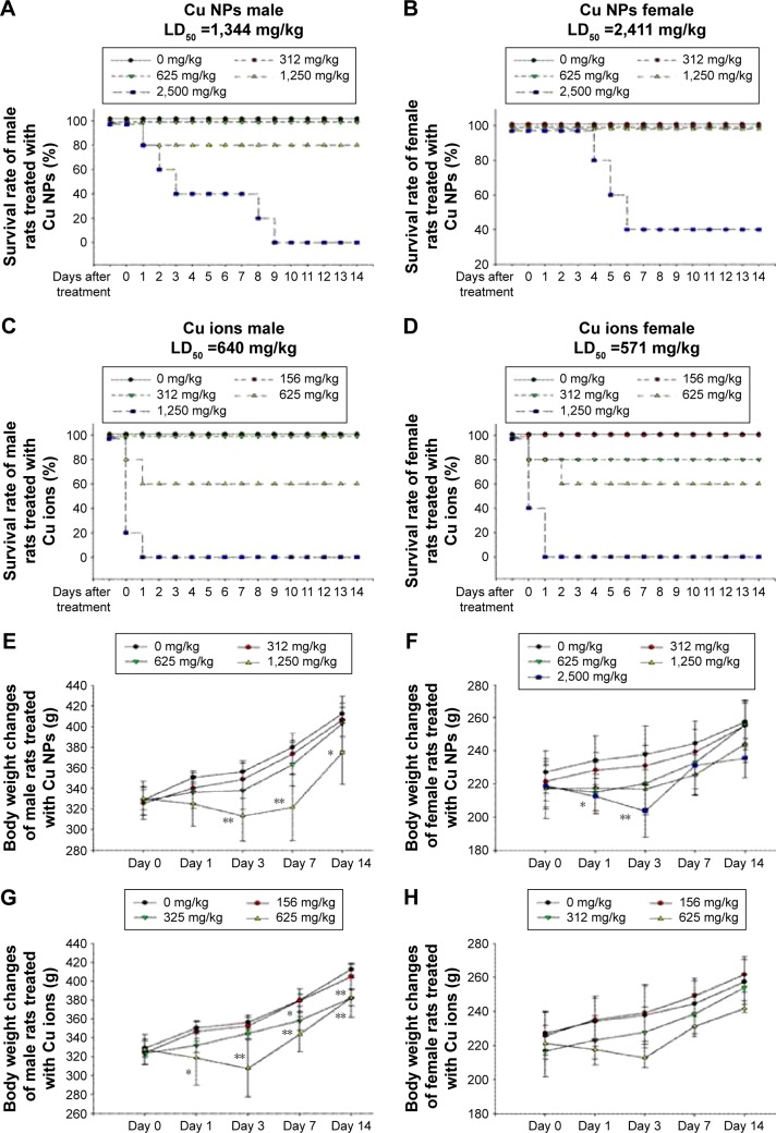
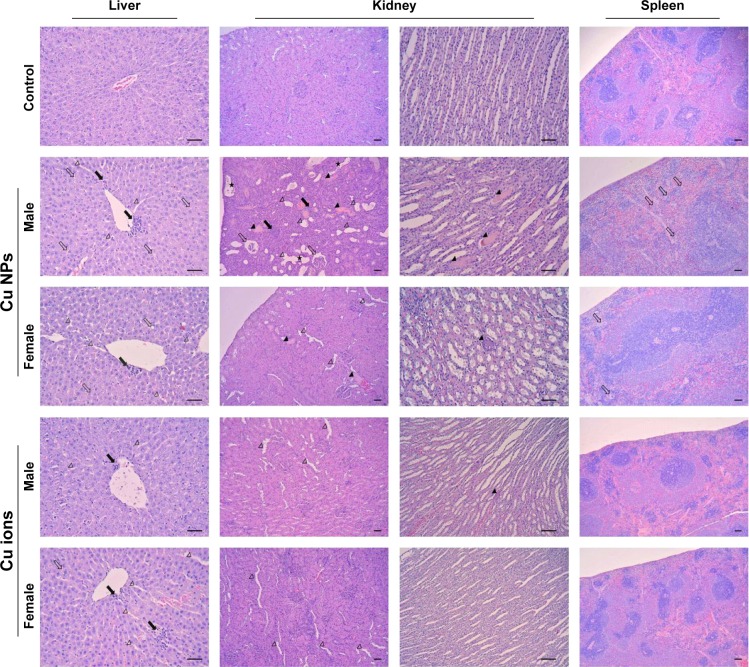
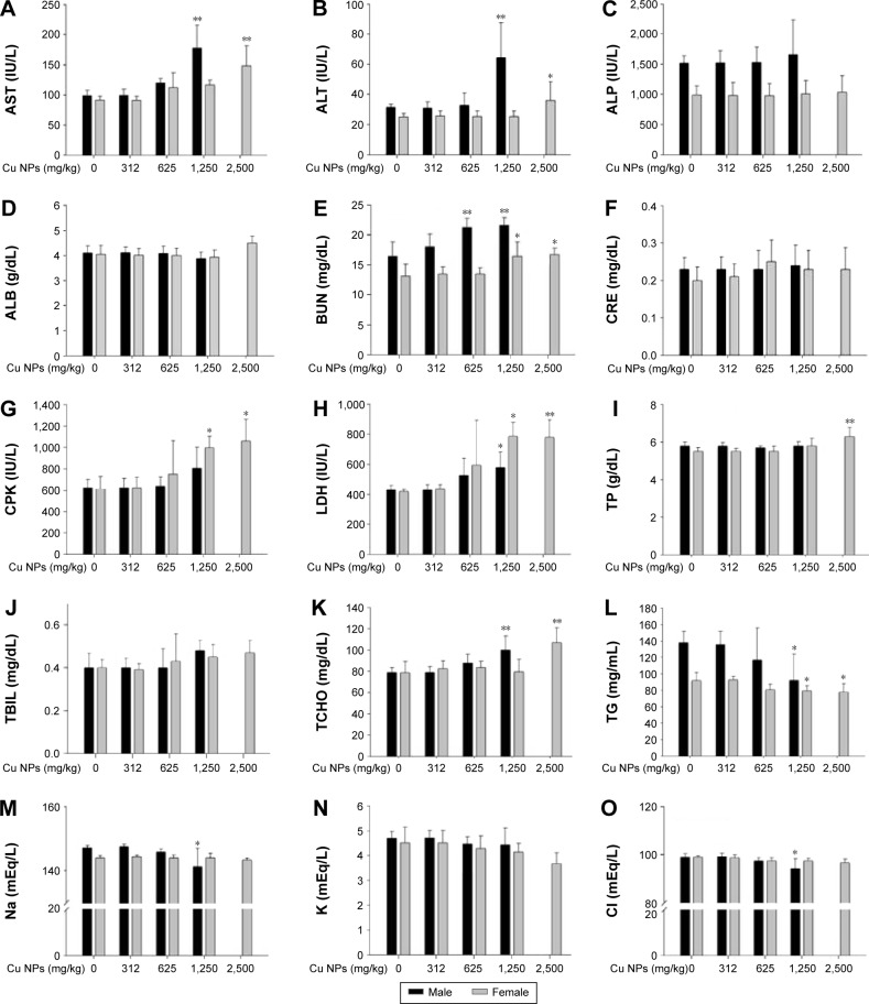
Despite widespread use and prospective biomedical applications of copper nanoparticles (Cu NPs), their biosafety issues and kinetics remain unclear. Thus, the aim of this study was to compare the detailed in vivo toxicity of Cu NPs and cupric ions (CuCl2; Cu ions) after a single oral dose. We determined the physicochemical characteristics of Cu NPs, including morphology, hydrodynamic size, zeta potential, and dissolution in gastric (pH 1.5), vehicle (pH 6.5), and intestinal (pH 7.8) conditions. We also evaluated the kinetics of Cu following a single equivalent dose (500 mg/kg) of Cu NPs and Cu ions. Cu NPs had highest dissolution (84.5%) only in gastric conditions when compared with complete dissolution of Cu ions under various physiological milieus. Kinetic analysis revealed that highest Cu levels in blood and tested organs of Cu NP-treated rats were 15%-25% lower than that of Cu ions. Similar to the case of Cu ions, Cu levels in the tested organs (especially liver, kidney, and spleen) of Cu NP-treated rats increased significantly when compared with the vehicle control. However, delay in reaching the highest level and biopersistence of Cu were observed in the blood and tested organs of Cu NP-treated rats compared with Cu ions. Extremely high levels of Cu in feces indicated that unabsorbed Cu NPs or absorbed Cu ions were predominantly eliminated through liver/feces. Cu NPs exerted apparent toxicological effects at higher dose levels compared with Cu ions and showed sex-dependent differences in mortality, biochemistry, and histopathology. Liver, kidney, and spleen were the major organs affected by Cu NPs. Collectively, the toxicity and kinetics of Cu NPs are most likely influenced by the release of Cu dissociated from Cu NPs under physiological conditions.

摘要

尽管铜纳米颗粒(Cu NPs)已被广泛应用并具有潜在的生物医学应用前景,但其生物安全性问题和动力学仍不清楚。因此,本研究的目的是比较单次口服剂量后Cu NPs和铜离子(CuCl2;Cu离子)在体内的详细毒性。我们测定了Cu NPs的物理化学特性,包括形态、流体动力学尺寸、zeta电位以及在胃(pH 1.5)、载体(pH 6.5)和肠道(pH 7.8)条件下的溶解性。我们还评估了单次等效剂量(500 mg/kg)的Cu NPs和Cu离子后铜的动力学。与在各种生理环境下完全溶解的Cu离子相比,Cu NPs仅在胃条件下具有最高的溶解性(84.5%)。动力学分析表明,Cu NP处理大鼠的血液和受试器官中的最高铜水平比Cu离子低15%-25%。与Cu离子的情况类似,与载体对照组相比,Cu NP处理大鼠的受试器官(尤其是肝脏、肾脏和脾脏)中的铜水平显著升高。然而,与Cu离子相比,在Cu NP处理大鼠的血液和受试器官中观察到铜达到最高水平的延迟和生物持久性。粪便中极高水平的铜表明未吸收的Cu NPs或吸收的Cu离子主要通过肝脏/粪便排出。与Cu离子相比,Cu NPs在较高剂量水平下表现出明显的毒理学效应,并且在死亡率、生物化学和组织病理学方面表现出性别依赖性差异。肝脏、肾脏和脾脏是受Cu NPs影响的主要器官。总体而言,Cu NPs的毒性和动力学很可能受到生理条件下从Cu NPs解离的铜释放的影响。

https://cdn.ncbi.nlm.nih.gov/pmc/blobs/aa24/4913985/5aa58ac96bc9/ijn-11-2883Fig8.jpg
https://cdn.ncbi.nlm.nih.gov/pmc/blobs/aa24/4913985/55b8ae0a1cec/ijn-11-2883Fig1.jpg
https://cdn.ncbi.nlm.nih.gov/pmc/blobs/aa24/4913985/4786c9c474d3/ijn-11-2883Fig2.jpg
https://cdn.ncbi.nlm.nih.gov/pmc/blobs/aa24/4913985/52f15c0fa8d7/ijn-11-2883Fig3.jpg
https://cdn.ncbi.nlm.nih.gov/pmc/blobs/aa24/4913985/777f4a2d50bd/ijn-11-2883Fig4.jpg
https://cdn.ncbi.nlm.nih.gov/pmc/blobs/aa24/4913985/d6a0ce4ae329/ijn-11-2883Fig5.jpg
https://cdn.ncbi.nlm.nih.gov/pmc/blobs/aa24/4913985/676a4f6b2e19/ijn-11-2883Fig6.jpg
https://cdn.ncbi.nlm.nih.gov/pmc/blobs/aa24/4913985/5d7edff9d5bd/ijn-11-2883Fig7.jpg
https://cdn.ncbi.nlm.nih.gov/pmc/blobs/aa24/4913985/5aa58ac96bc9/ijn-11-2883Fig8.jpg

相似文献

1
Comparative toxicity and biodistribution of copper nanoparticles and cupric ions in rats.大鼠体内铜纳米颗粒和铜离子的比较毒性及生物分布
Int J Nanomedicine. 2016 Jun 16;11:2883-900. doi: 10.2147/IJN.S106346. eCollection 2016.
2
Comparative toxicity and biodistribution assessments in rats following subchronic oral exposure to copper nanoparticles and microparticles.大鼠亚慢性经口暴露于铜纳米颗粒和微米颗粒后的比较毒性及生物分布评估。
Part Fibre Toxicol. 2016 Oct 28;13(1):56. doi: 10.1186/s12989-016-0169-x.
3
Acute toxicity and biodistribution of different sized copper nano-particles in rats after oral administration.经口给予不同粒径铜纳米粒子后大鼠的急性毒性和生物分布。
Mater Sci Eng C Mater Biol Appl. 2018 Dec 1;93:649-663. doi: 10.1016/j.msec.2018.08.032. Epub 2018 Aug 14.
4
Toxicity of different-sized copper nano- and submicron particles and their shed copper ions to zebrafish embryos.不同粒径的铜纳米和亚微米颗粒及其释放的铜离子对斑马鱼胚胎的毒性。
Environ Toxicol Chem. 2014 Aug;33(8):1774-82. doi: 10.1002/etc.2615. Epub 2014 Jun 20.
5
Effects and mechanisms of sub-chronic exposure to copper nanoparticles on renal cytochrome P450 enzymes in rats.亚慢性暴露于铜纳米颗粒对大鼠肾细胞色素 P450 酶的影响及其作用机制。
Environ Toxicol Pharmacol. 2018 Oct;63:135-146. doi: 10.1016/j.etap.2018.08.004. Epub 2018 Aug 7.
6
Dissolution and Persistence of Copper-Based Nanomaterials in Undersaturated Solutions with Respect to Cupric Solid Phases.铜基纳米材料在相对于铜固相不饱和溶液中的溶解和持久行为。
Environ Sci Technol. 2016 Jul 5;50(13):6772-81. doi: 10.1021/acs.est.5b04719. Epub 2016 Jan 13.
7
Synthesis methods influence characteristics, behaviour and toxicity of bare CuO NPs compared to bulk CuO and ionic Cu after in vitro exposure of Ruditapes philippinarum hemocytes.与体外暴露于菲律宾蛤仔血细胞的体相 CuO 和离子态 Cu 相比,合成方法会影响裸 CuO NPs 的特性、行为和毒性。
Aquat Toxicol. 2018 Jun;199:285-295. doi: 10.1016/j.aquatox.2018.04.007. Epub 2018 Apr 17.
8
Copper status of exposed microorganisms influences susceptibility to metallic nanoparticles.暴露微生物的铜状态会影响其对金属纳米颗粒的敏感性。
Environ Toxicol Chem. 2016 May;35(5):1148-58. doi: 10.1002/etc.3254. Epub 2016 Mar 9.
9
intestinal toxicity of copper oxide nanoparticles in rat and human cell models.氧化铜纳米粒子在大鼠和人体细胞模型中的肠道毒性。
Nanotoxicology. 2019 Aug;13(6):795-811. doi: 10.1080/17435390.2019.1578428. Epub 2019 Apr 2.
10
Copper nanoparticles lead to reproductive dysfunction by affecting key enzymes of ovarian hormone synthesis and metabolism in female rats.铜纳米颗粒通过影响雌性大鼠卵巢激素合成和代谢的关键酶导致生殖功能障碍。
Ecotoxicol Environ Saf. 2023 Apr 1;254:114704. doi: 10.1016/j.ecoenv.2023.114704. Epub 2023 Mar 8.

引用本文的文献

1
Exposure to Fine Particulate Matter (PM2.5) and Heavy Metals During the Second Trimester of Pregnancy Increases the Risk of Preeclampsia and Eclampsia: An Analysis of National Health Insurance Claims Data from South Korea.孕期中期暴露于细颗粒物(PM2.5)和重金属会增加子痫前期和子痫的风险:来自韩国国民健康保险索赔数据的分析
Medicina (Kaunas). 2025 Jun 25;61(7):1146. doi: 10.3390/medicina61071146.
2
Influence of capping agents on physicochemical properties and leukemic cytotoxicity of copper oxide nanoparticles biosynthesized using Caesalpinia sappan extract.封端剂对使用苏木提取物生物合成的氧化铜纳米颗粒的物理化学性质和白血病细胞毒性的影响。
PLoS One. 2025 Jun 26;20(6):e0326791. doi: 10.1371/journal.pone.0326791. eCollection 2025.
3

本文引用的文献

1
Copper-based nanoparticles induce high toxicity in leukemic HL60 cells.铜基纳米颗粒对白血病HL60细胞具有高毒性。
Toxicol In Vitro. 2015 Oct;29(7):1711-9. doi: 10.1016/j.tiv.2015.05.020. Epub 2015 May 28.
2
Implications of protein corona on physico-chemical and biological properties of magnetic nanoparticles.蛋白质冠层对磁性纳米颗粒物理化学和生物学性质的影响。
Biomaterials. 2015 Apr;46:1-12. doi: 10.1016/j.biomaterials.2014.12.045. Epub 2015 Jan 15.
3
Acute exposure to ZnO nanoparticles induces autophagic immune cell death.
Cardiovascular Toxicity of Metal-Based Nanoparticles.金属基纳米颗粒的心血管毒性
Int J Mol Sci. 2025 Jun 17;26(12):5816. doi: 10.3390/ijms26125816.
4
Inulin Improves the Redox Response in Rats Fed a Diet Containing Recommended Copper Nanoparticle (CuNPs) Levels, While Pectin or Psyllium in Rats Receive Excessive CuNPs Levels in the Diet.菊粉可改善喂食含推荐水平铜纳米颗粒(CuNPs)日粮的大鼠的氧化还原反应,而大鼠日粮中的果胶或车前子壳会摄入过量的CuNPs。
Antioxidants (Basel). 2025 Jun 8;14(6):695. doi: 10.3390/antiox14060695.
5
CuO-NPs Induce Apoptosis and Functional Impairment in BV2 Cells Through the CSF-1R/PLCγ2/ERK/Nrf2 Pathway.氧化铜纳米颗粒通过CSF-1R/PLCγ2/ERK/Nrf2途径诱导BV2细胞凋亡和功能损伤。
Toxics. 2025 Mar 21;13(4):231. doi: 10.3390/toxics13040231.
6
Acute brain injury and nanomedicine: sex as a biological variable.急性脑损伤与纳米医学:性别作为一种生物学变量
Front Biomater Sci. 2024;3. doi: 10.3389/fbiom.2024.1348165. Epub 2024 Feb 2.
7
Skin repair and infection control in diabetic, obese mice using bioactive laser-activated sealants.利用生物活性激光激活密封剂修复糖尿病肥胖小鼠皮肤并控制感染。
Biomaterials. 2024 Dec;311:122668. doi: 10.1016/j.biomaterials.2024.122668. Epub 2024 Jun 14.
8
Electrospun Nanofiber Scaffolds Loaded with Metal-Based Nanoparticles for Wound Healing.负载金属基纳米颗粒的电纺纳米纤维支架用于伤口愈合
Polymers (Basel). 2023 Dec 20;16(1):24. doi: 10.3390/polym16010024.
9
Conversion of senescent cartilage into a pro-chondrogenic microenvironment with antibody-functionalized copper sulfate nanoparticles for efficient osteoarthritis therapy.用抗体功能化的硫酸铜纳米粒子将衰老的软骨转化为促软骨形成的微环境,用于高效的骨关节炎治疗。
J Nanobiotechnology. 2023 Aug 8;21(1):258. doi: 10.1186/s12951-023-02036-5.
10
Efficacy of zinc oxide and copper oxide nanoparticles on virulence genes of avian pathogenic E. coli (APEC) in broilers.氧化锌和氧化铜纳米粒子对肉鸡致病性大肠杆菌(APEC)毒力基因的影响。
BMC Vet Res. 2023 Aug 4;19(1):108. doi: 10.1186/s12917-023-03643-y.
急性暴露于氧化锌纳米颗粒会诱导自噬免疫细胞死亡。
Nanotoxicology. 2015;9(6):737-48. doi: 10.3109/17435390.2014.974709. Epub 2014 Nov 7.
4
Studies on the toxicity and distribution of indium compounds according to particle size in sprague-dawley rats.根据粒径研究铟化合物在 Sprague-Dawley 大鼠中的毒性和分布。
Toxicol Res. 2014 Mar;30(1):55-63. doi: 10.5487/TR.2014.30.1.055.
5
Bioactivity, mechanism of action, and cytotoxicity of copper-based nanoparticles: a review.铜基纳米颗粒的生物活性、作用机制及细胞毒性:综述
Appl Microbiol Biotechnol. 2014 Feb;98(3):1001-9. doi: 10.1007/s00253-013-5422-8. Epub 2013 Dec 5.
6
Comparison of toxicity and deposition of nano-sized carbon black aerosol prepared with or without dispersing sonication.比较经或未经超声分散处理制备的纳米级炭黑气溶胶的毒性和沉积。
Toxicol Res. 2013 Jun;29(2):121-7. doi: 10.5487/TR.2013.29.2.121.
7
Nanoparticle toxicity by the gastrointestinal route: evidence and knowledge gaps.纳米颗粒经胃肠道途径的毒性:证据与知识空白
Int J Biomed Nanosci Nanotechnol. 2013;3(1-2). doi: 10.1504/IJBNN.2013.054515.
8
A systematic in-vivo toxicity evaluation of nanophosphor particles via zebrafish models.通过斑马鱼模型对纳米荧光粉颗粒进行系统的体内毒性评价。
Biomaterials. 2014 Jan;35(1):440-9. doi: 10.1016/j.biomaterials.2013.09.054. Epub 2013 Oct 4.
9
Copper oxide nanoparticles induced mitochondria mediated apoptosis in human hepatocarcinoma cells.氧化铜纳米颗粒诱导人肝癌细胞线粒体介导的细胞凋亡。
PLoS One. 2013 Aug 5;8(8):e69534. doi: 10.1371/journal.pone.0069534. Print 2013.
10
Toxicity of Ag, CuO and ZnO nanoparticles to selected environmentally relevant test organisms and mammalian cells in vitro: a critical review.纳米银、氧化铜和氧化锌颗粒对选定的环境相关测试生物和哺乳动物细胞的体外毒性:一项批判性综述。
Arch Toxicol. 2013 Jul;87(7):1181-200. doi: 10.1007/s00204-013-1079-4. Epub 2013 Jun 1.