• 文献检索
  • 文档翻译
  • 深度研究
  • 学术资讯
  • Suppr Zotero 插件Zotero 插件
  • 邀请有礼
  • 套餐&价格
  • 历史记录
应用&插件
Suppr Zotero 插件Zotero 插件浏览器插件Mac 客户端Windows 客户端微信小程序
定价
高级版会员购买积分包购买API积分包
服务
文献检索文档翻译深度研究API 文档MCP 服务
关于我们
关于 Suppr公司介绍联系我们用户协议隐私条款
关注我们

Suppr 超能文献

核心技术专利:CN118964589B侵权必究
粤ICP备2023148730 号-1Suppr @ 2026

文献检索

告别复杂PubMed语法,用中文像聊天一样搜索,搜遍4000万医学文献。AI智能推荐,让科研检索更轻松。

立即免费搜索

文件翻译

保留排版,准确专业,支持PDF/Word/PPT等文件格式,支持 12+语言互译。

免费翻译文档

深度研究

AI帮你快速写综述,25分钟生成高质量综述,智能提取关键信息,辅助科研写作。

立即免费体验

纤连蛋白接枝钛牙种植体:一项体内研究。

Fibronectin-Grafted Titanium Dental Implants: An In Vivo Study.

作者信息

Chang Yu-Chi, Ho Kuo-Ning, Feng Sheng-Wei, Huang Haw-Ming, Chang Chia-Hsun, Lin Che-Tong, Teng Nai-Chia, Pan Yu Hwa, Chang Wei-Jen

机构信息

School of Dentistry, College of Oral Medicine, Taipei Medical University, Taipei 110, Taiwan.

School of Dentistry, College of Oral Medicine, Taipei Medical University, Taipei 110, Taiwan; Graduate Institute of Biomedical Materials & Tissue Engineering, College of Oral Medicine, Taipei Medical University, Taipei 110, Taiwan.

出版信息

Biomed Res Int. 2016;2016:2414809. doi: 10.1155/2016/2414809. Epub 2016 Jun 6.

DOI:10.1155/2016/2414809
PMID:27366739
原文链接:https://pmc.ncbi.nlm.nih.gov/articles/PMC4913050/
Abstract

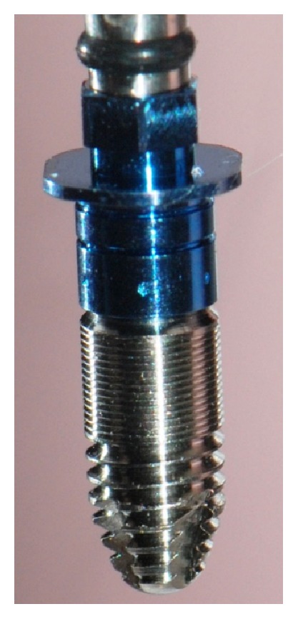
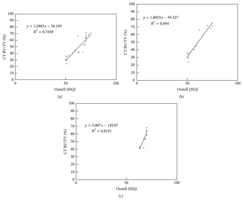
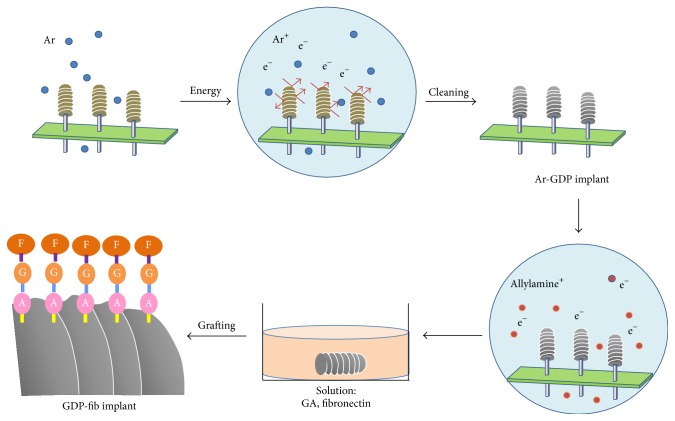
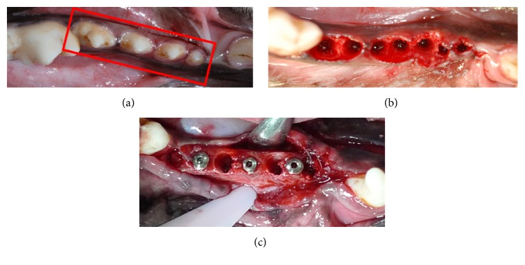
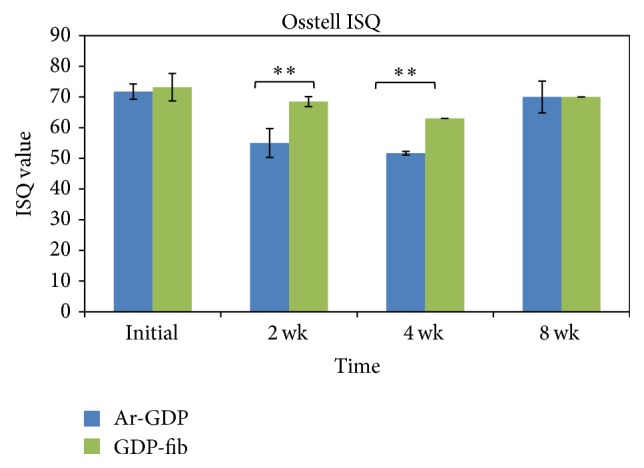
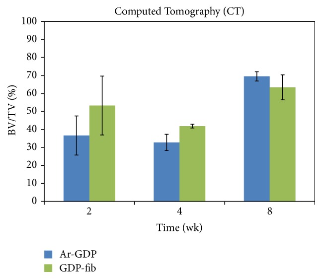
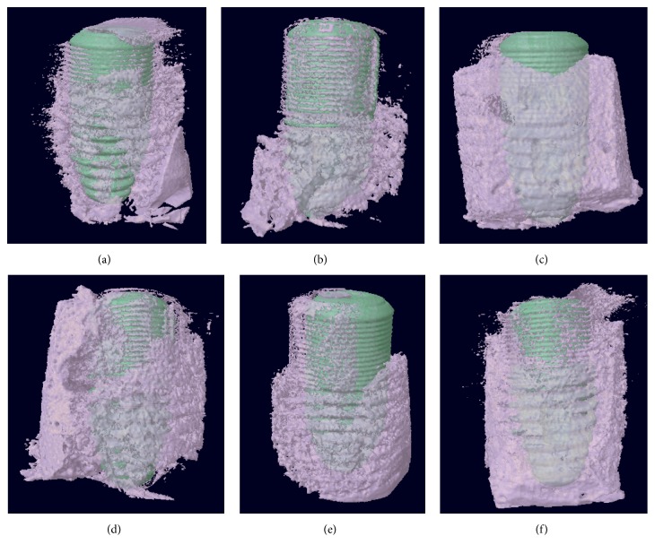
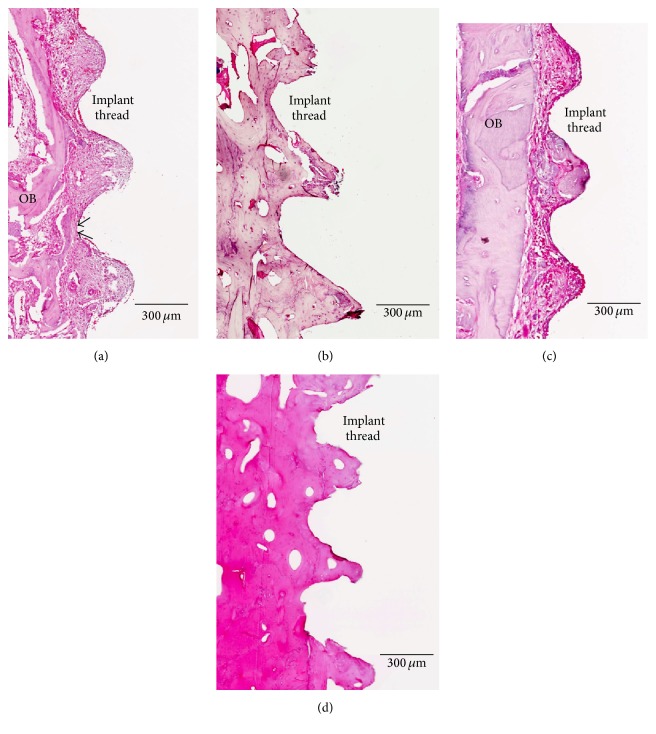
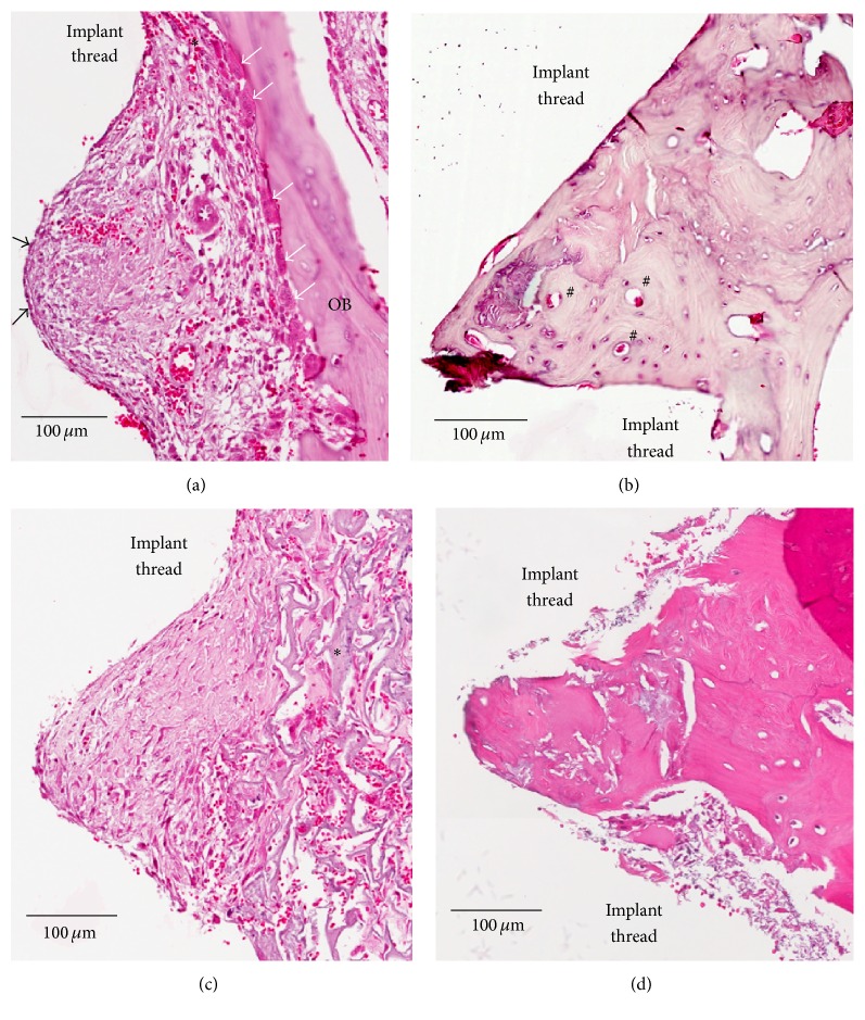
Modification of the physiochemical properties of titanium surfaces using glow discharge plasma (GDP) and fibronectin coating has been shown to enhance the surface hydrophilicity, surface roughness, cell adhesion, migration, and proliferation. This in vivo study aimed to evaluate the bone integration efficacy of a biologically modified implant surface. Two different surface-modified implants (Ar-GDP and GDP-fib) were placed in the mandibular premolar area of six beagle dogs for 2-8 weeks. Three techniques [histologic evaluation, resonance frequency analysis (RFA), and microcomputed tomography (micro-CT) evaluation] were used to detect the implant stability and bone-implant contact. The implant stability quotient values of GDP-fib implants were significantly greater than the Ar-GDP implants at 2 and 4 weeks (P < 0.01). The bone volume/total volume ratio of GDP-fib implants was greater than the Ar-GDP implants in micro-CT evaluation. A high positive correlation was observed between RFA and micro-CT measurements. At 2 weeks, osteoblasts were seen to line the implant surface, and multinuclear osteoclasts could be seen on the surface of old parent bone. After 8 weeks, a majority of the space in the wound chamber appeared to be replaced by bone. Enhancement of the stability of biologically modified implants was proved by the results of RFA, micro-CT, and histological analysis. This enhanced stability may help fasten treatment and be clinically beneficial.

摘要

使用辉光放电等离子体(GDP)和纤连蛋白涂层对钛表面的物理化学性质进行改性,已被证明可增强表面亲水性、表面粗糙度、细胞粘附、迁移和增殖。本体内研究旨在评估生物改性种植体表面的骨整合效果。将两种不同表面改性的种植体(Ar-GDP和GDP-纤连蛋白)植入6只比格犬的下颌前磨牙区2至8周。使用三种技术[组织学评估、共振频率分析(RFA)和微型计算机断层扫描(micro-CT)评估]来检测种植体稳定性和骨-种植体接触情况。在2周和4周时,GDP-纤连蛋白种植体的种植体稳定性商值显著高于Ar-GDP种植体(P < 0.01)。在微型计算机断层扫描评估中,GDP-纤连蛋白种植体的骨体积/总体积比大于Ar-GDP种植体。在RFA和微型计算机断层扫描测量之间观察到高度正相关。在2周时,可见成骨细胞排列在种植体表面,在旧母骨表面可见多核破骨细胞。8周后,伤口腔内的大部分空间似乎被骨替代。RFA、微型计算机断层扫描和组织学分析结果证明了生物改性种植体稳定性的增强。这种增强的稳定性可能有助于加快治疗并具有临床益处。

https://cdn.ncbi.nlm.nih.gov/pmc/blobs/8399/4913050/069d764b0609/BMRI2016-2414809.009.jpg
https://cdn.ncbi.nlm.nih.gov/pmc/blobs/8399/4913050/e42a2e4803ec/BMRI2016-2414809.001.jpg
https://cdn.ncbi.nlm.nih.gov/pmc/blobs/8399/4913050/71f2876ddf02/BMRI2016-2414809.002.jpg
https://cdn.ncbi.nlm.nih.gov/pmc/blobs/8399/4913050/d584fd4ea159/BMRI2016-2414809.003.jpg
https://cdn.ncbi.nlm.nih.gov/pmc/blobs/8399/4913050/979eef63c780/BMRI2016-2414809.004.jpg
https://cdn.ncbi.nlm.nih.gov/pmc/blobs/8399/4913050/5d271444b2b8/BMRI2016-2414809.005.jpg
https://cdn.ncbi.nlm.nih.gov/pmc/blobs/8399/4913050/b9c0ea1a0a64/BMRI2016-2414809.006.jpg
https://cdn.ncbi.nlm.nih.gov/pmc/blobs/8399/4913050/7aa554097c17/BMRI2016-2414809.007.jpg
https://cdn.ncbi.nlm.nih.gov/pmc/blobs/8399/4913050/c56e07f97370/BMRI2016-2414809.008.jpg
https://cdn.ncbi.nlm.nih.gov/pmc/blobs/8399/4913050/069d764b0609/BMRI2016-2414809.009.jpg

相似文献

1
Fibronectin-Grafted Titanium Dental Implants: An In Vivo Study.纤连蛋白接枝钛牙种植体:一项体内研究。
Biomed Res Int. 2016;2016:2414809. doi: 10.1155/2016/2414809. Epub 2016 Jun 6.
2
Effect of implant surface properties on peri-implant bone healing: implant stability and microcomputed tomographic analysis.种植体表面特性对种植体周围骨愈合的影响:种植体稳定性和微计算机断层扫描分析。
Int J Oral Maxillofac Implants. 2012 Jan-Feb;27(1):77-83.
3
Surface analysis of titanium biological modification with glow discharge.辉光放电对钛进行生物改性的表面分析
Clin Implant Dent Relat Res. 2015 Jun;17(3):469-75. doi: 10.1111/cid.12141. Epub 2013 Aug 27.
4
The role of titanium implant surface modification with hydroxyapatite nanoparticles in progressive early bone-implant fixation in vivo.羟基磷灰石纳米颗粒改性钛种植体表面在体内渐进性早期骨-种植体固定中的作用。
Int J Oral Maxillofac Implants. 2009 Sep-Oct;24(5):808-16.
5
In vivo monitoring of the bone healing process around different titanium alloy implant surfaces placed into fresh extraction sockets.在体监测不同钛合金种植体表面植入新鲜拔牙窝周围的骨愈合过程。
J Dent. 2012 Apr;40(4):338-46. doi: 10.1016/j.jdent.2012.01.010. Epub 2012 Jan 28.
6
Biological response to titanium implants coated with nanocrystals calcium phosphate or type 1 collagen in a dog model.纳米晶磷酸钙或 1 型胶原涂层钛植入物在犬模型中的生物学反应。
Clin Oral Implants Res. 2013 May;24(5):475-83. doi: 10.1111/j.1600-0501.2011.02409.x. Epub 2012 Jan 26.
7
In Vitro Analysis of Fibronectin-Modified Titanium Surfaces.纤连蛋白修饰钛表面的体外分析
PLoS One. 2016 Jan 5;11(1):e0146219. doi: 10.1371/journal.pone.0146219. eCollection 2016.
8
Anodization increases early integration of Osstem implants in sheep femurs.阳极氧化可促进奥齿泰种植体在绵羊股骨中的早期整合。
Ann R Australas Coll Dent Surg. 2008 Jun;19:152-6.
9
Resolution, sensitivity, and in vivo application of high-resolution computed tomography for titanium-coated polymethyl methacrylate (PMMA) dental implants.钛涂层聚甲基丙烯酸甲酯(PMMA)牙科植入物的高分辨率计算机断层扫描的分辨率、灵敏度及体内应用
Clin Oral Implants Res. 2014 Mar;25(3):359-365. doi: 10.1111/clr.12128. Epub 2013 Feb 20.
10
Effects of a cell adhesion molecule coating on the blasted surface of titanium implants on bone healing in the rabbit femur.细胞粘附分子涂层对兔股骨钛植入物喷砂表面骨愈合的影响。
Int J Oral Maxillofac Implants. 2007 Jul-Aug;22(4):533-41.

引用本文的文献

1
Assessment of Low-Dose rhBMP-2 and Vacuum Plasma Treatments on Titanium Implants for Osseointegration and Bone Regeneration.低剂量重组人骨形态发生蛋白-2和真空等离子体处理对钛种植体骨整合和骨再生的评估
Materials (Basel). 2025 Jul 30;18(15):3582. doi: 10.3390/ma18153582.
2
Biomolecule-functionalized dental implant surfaces: Towards augmenting soft tissue integration.生物分子功能化的牙种植体表面:促进软组织整合
Bioact Mater. 2025 Jul 26;53:540-590. doi: 10.1016/j.bioactmat.2025.07.005. eCollection 2025 Nov.
3
Construction of functional surfaces for dental implants to enhance osseointegration.

本文引用的文献

1
In Vitro Analysis of Fibronectin-Modified Titanium Surfaces.纤连蛋白修饰钛表面的体外分析
PLoS One. 2016 Jan 5;11(1):e0146219. doi: 10.1371/journal.pone.0146219. eCollection 2016.
2
Surface analysis of titanium biological modification with glow discharge.辉光放电对钛进行生物改性的表面分析
Clin Implant Dent Relat Res. 2015 Jun;17(3):469-75. doi: 10.1111/cid.12141. Epub 2013 Aug 27.
3
Heat and radiofrequency plasma glow discharge pretreatment of a titanium alloy promote bone formation and osseointegration.热和射频等离子体辉光放电预处理钛合金可促进骨形成和骨整合。
用于牙种植体的功能性表面构建以增强骨整合。
Front Bioeng Biotechnol. 2023 Nov 14;11:1320307. doi: 10.3389/fbioe.2023.1320307. eCollection 2023.
4
3D-Printed PLA Scaffold with Fibronectin Enhances In Vitro Osteogenesis.含纤连蛋白的3D打印聚乳酸支架增强体外成骨作用。
Polymers (Basel). 2023 Jun 8;15(12):2619. doi: 10.3390/polym15122619.
5
Guiding Fibroblast Activation Using an RGD-Mutated Heparin Binding II Fragment of Fibronectin for Gingival Titanium Integration.利用纤维连接蛋白 RGD 突变肝素结合 II 片段引导成纤维细胞活化以增强牙龈钛整合。
Adv Healthc Mater. 2023 Aug;12(21):e2203307. doi: 10.1002/adhm.202203307. Epub 2023 May 10.
6
Fibronectin and Its Applications in Dentistry and Periodontics: A Cell Behaviour Conditioner.纤连蛋白及其在牙科学和牙周病学中的应用:一种细胞行为调节剂。
Cureus. 2022 Oct 26;14(10):e30702. doi: 10.7759/cureus.30702. eCollection 2022 Oct.
7
Titanium Dental Implants: An Overview of Applied Nanobiotechnology to Improve Biocompatibility and Prevent Infections.钛牙种植体:应用纳米生物技术改善生物相容性和预防感染的概述
Materials (Basel). 2022 Apr 27;15(9):3150. doi: 10.3390/ma15093150.
8
Modification of implant surfaces to stimulate mesenchymal cell activation.对植入物表面进行改性以刺激间充质细胞活化。
Bull Natl Res Cent. 2022;46(1):52. doi: 10.1186/s42269-022-00743-x. Epub 2022 Mar 4.
9
Biochemical Modification of Titanium Oral Implants: Evidence from In Vivo Studies.钛口腔种植体的生化改性:来自体内研究的证据
Materials (Basel). 2021 May 24;14(11):2798. doi: 10.3390/ma14112798.
10
Simple application of adipose-derived stem cell-derived extracellular vesicles coating enhances cytocompatibility and osteoinductivity of titanium implant.脂肪来源干细胞衍生的细胞外囊泡涂层的简单应用可增强钛植入物的细胞相容性和骨诱导性。
Regen Biomater. 2020 Dec 3;8(1):rbaa038. doi: 10.1093/rb/rbaa038. eCollection 2021 Feb 1.
J Cell Biochem. 2013 Oct;114(10):2363-74. doi: 10.1002/jcb.24585.
4
Influence on early osseointegration of dental implants installed with two different drilling protocols: a histomorphometric study in rabbit.两种不同钻备 protocols 对种植体早期骨整合影响的组织形态计量学研究:兔。
Clin Oral Implants Res. 2011 Jan;22(1):92-9. doi: 10.1111/j.1600-0501.2010.02009.x. Epub 2010 Oct 6.
5
The quantitative assessment of peri-implant bone responses using histomorphometry and micro-computed tomography.使用组织形态计量学和显微计算机断层扫描对种植体周围骨反应进行定量评估。
Biomaterials. 2009 Sep;30(27):4539-49. doi: 10.1016/j.biomaterials.2009.05.017. Epub 2009 Jun 4.
6
Potential of chemically modified hydrophilic surface characteristics to support tissue integration of titanium dental implants.化学修饰亲水性表面特性对支持钛牙种植体组织整合的潜力。
J Biomed Mater Res B Appl Biomater. 2009 Feb;88(2):544-57. doi: 10.1002/jbm.b.31233.
7
Type I collagen grafting on titanium surfaces using low-temperature glow discharge.使用低温辉光放电在钛表面进行I型胶原移植。
Dent Mater J. 2008 May;27(3):340-6. doi: 10.4012/dmj.27.340.
8
The effect of integrin-specific bioactive coatings on tissue healing and implant osseointegration.整合素特异性生物活性涂层对组织愈合和种植体骨整合的影响。
Biomaterials. 2008 Jul;29(19):2849-57. doi: 10.1016/j.biomaterials.2008.03.036. Epub 2008 Apr 11.
9
Enhanced implant stability with a chemically modified SLA surface: a randomized pilot study.通过化学改性的SLA表面增强种植体稳定性:一项随机试点研究。
Int J Oral Maxillofac Implants. 2007 Sep-Oct;22(5):755-60.
10
Enhanced osseointegration by the chemotactic activity of plasma fibronectin for cellular fibronectin positive cells.通过血浆纤连蛋白对细胞纤连蛋白阳性细胞的趋化活性增强骨结合。
Biomaterials. 2007 Aug;28(24):3469-77. doi: 10.1016/j.biomaterials.2007.04.029. Epub 2007 May 3.