Costagliola Alessandro, Piegari Giuseppe, Otrocka-Domagala Iwona, Ciccarelli Davide, Iovane Valentina, Oliva Gaetano, Russo Valeria, Rinaldi Laura, Papparella Serenella, Paciello Orlando
Unit of Pathology, Department of Veterinary Medicine and Animal Productions, University of Naples Federico II, 80137 Naples, Italy.
Department of Pathological Anatomy, Faculty of Veterinary Medicine, Warmia and Mazury University in Olsztyn, 10-701 Olsztyn, Poland.
Biomed Res Int. 2016;2016:8016186. doi: 10.1155/2016/8016186. Epub 2016 Jun 19.
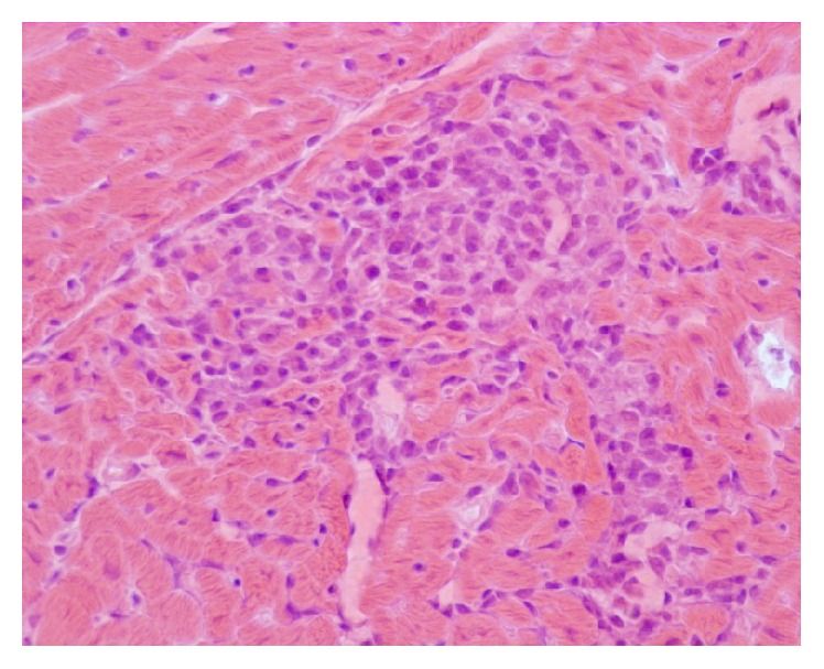
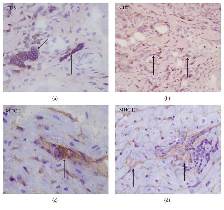
Myocarditis associated with infectious diseases may occur in dogs, including those caused by the protozoa Neospora caninum, Trypanosoma cruzi, Babesia canis, and Hepatozoon canis. However, although cardiac disease due to Leishmania infection has also been documented, the immunopathological features of myocarditis have not been reported so far. The aim of this study was to examine the types of cellular infiltrates and expression of MHC classes I and II in myocardial samples obtained at necropsy from 15 dogs with an established intravitam diagnosis of visceral leishmaniasis. Pathological features of myocardium were characterized by hyaline degeneration of cardiomyocytes, necrosis, and infiltration of mononuclear inflammatory cells consisting of lymphocytes and macrophages, sometimes with perivascular pattern; fibrosis was also present in various degrees. Immunophenotyping of inflammatory cells was performed by immunohistochemistry on cryostat sections obtained from the heart of the infected dogs. The predominant leukocyte population was CD8+ with a fewer number of CD4+ cells. Many cardiomyocytes expressed MHC classes I and II on the sarcolemma. Leishmania amastigote forms were not detected within macrophages or any other cell of the examined samples. Our study provided evidence that myocarditis in canine visceral leishmaniasis might be related to immunological alterations associated with Leishmania infection.
与传染病相关的心肌炎可能发生在犬类中,包括由犬新孢子虫、克氏锥虫、犬巴贝斯虫和犬肝簇虫等原生动物引起的心肌炎。然而,尽管利什曼原虫感染导致的心脏疾病也有文献记载,但迄今为止,心肌炎的免疫病理学特征尚未见报道。本研究的目的是检查从15只生前确诊为内脏利什曼病的犬尸检获得的心肌样本中的细胞浸润类型以及MHC I类和II类的表达。心肌的病理特征表现为心肌细胞玻璃样变性、坏死,以及由淋巴细胞和巨噬细胞组成的单核炎性细胞浸润,有时呈血管周围分布模式;不同程度的纤维化也存在。通过对感染犬心脏的冷冻切片进行免疫组织化学,对炎性细胞进行免疫表型分析。主要的白细胞群体是CD8 +,CD4 +细胞数量较少。许多心肌细胞在肌膜上表达MHC I类和II类。在所检查样本的巨噬细胞或任何其他细胞内未检测到利什曼原虫无鞭毛体形式。我们的研究提供了证据,表明犬内脏利什曼病中的心肌炎可能与利什曼原虫感染相关的免疫改变有关。