Shekhani Mohammed Talha, Forde Toni S, Adilbayeva Altynai, Ramez Mohamed, Myngbay Askhat, Bexeitov Yergali, Lindner Volkhard, Adarichev Vyacheslav A
Albert Einstein College of Medicine, Departments of Medicine (Division of Rheumatology) and Microbiology & Immunology, Bronx, NY, 10461, USA.
National Laboratory Astana, Astana, 010000, Kazakhstan.
Arthritis Res Ther. 2016 Jul 19;18:171. doi: 10.1186/s13075-016-1067-1.
The formation of destructive hypercellular pannus is critical to joint damage in rheumatoid arthritis (RA). The collagen triple helix repeat containing 1 (CTHRC1) protein expressed by activated stromal cells of diverse origin has previously been implicated in tissue remodeling and carcinogenesis. We recently discovered that the synovial Cthrc1 mRNA directly correlates with arthritis severity in mice. This study characterizes the role of CTHRC1 in arthritic pannus formation.
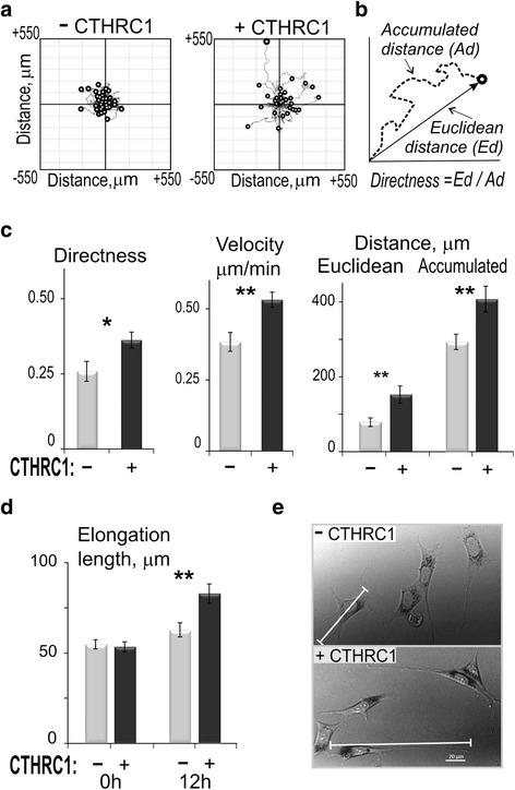
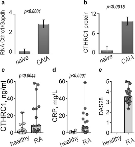
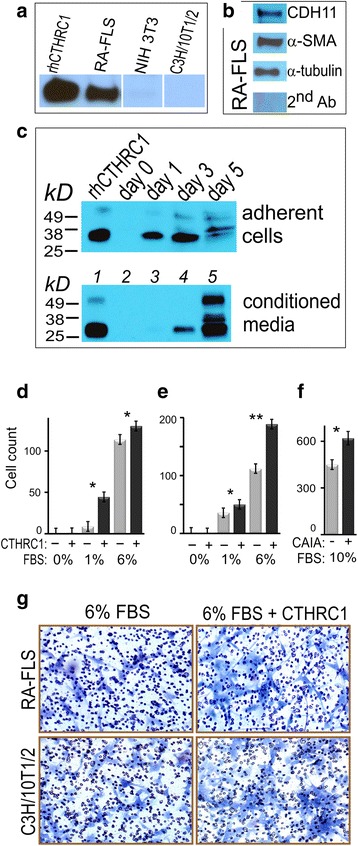
Synovial joints of mice with collagen antibody-induced arthritis (CAIA) and human RA-fibroblast-like synoviocytes (FLS) were immunostained for CTHRC1, FLS and macrophage-specific markers. CTHRC1 levels in plasma from patients with RA were measured using sandwich ELISA. The migratory response of fibroblasts was studied with a transwell migration assay and time-lapse microscopy. Velocity and directness of cell migration was analyzed by recording the trajectories of cells treated with rhCTHRC1.
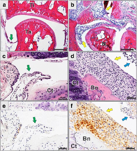
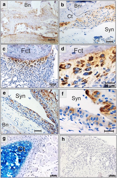
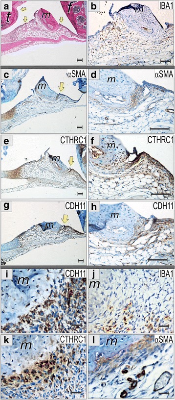
Immunohistochemical analysis of normal and inflamed synovium revealed highly inducible expression of CTHRC1 in arthritis (10.9-fold). At the tissue level, CTHRC1-expressing cells occupied the same niche as large fibroblast-like cells positive for α-smooth muscle actin (α-SMA) and cadherin 11 (CDH11). CTHRC1 was produced by activated FLS predominantly located at the synovial intimal lining and at the bone-pannus interface. Cultured RA-FLS expressed CDH11, α-SMA, and CTHRC1. Upon treatment with exogenous rhCTHRC1, embryonic fibroblasts and RA-FLS significantly increased migration velocity, directness, and cell length along the front-tail axis (1.4-fold, p < 0.01).
CTHRC1 was established as a novel marker of activated synoviocytes in murine experimental arthritis and RA. The pro-migratory effect of CTHRC1 on synoviocytes is considered one of the mechanisms promoting hypercellularity of the arthritic pannus.
破坏性的细胞增多性血管翳的形成对类风湿关节炎(RA)的关节损伤至关重要。先前已表明,多种来源的活化基质细胞表达的含胶原三螺旋重复序列1(CTHRC1)蛋白与组织重塑和致癌作用有关。我们最近发现,滑膜Cthrc1 mRNA与小鼠关节炎严重程度直接相关。本研究旨在阐明CTHRC1在关节炎性血管翳形成中的作用。
对胶原抗体诱导的关节炎(CAIA)小鼠的滑膜关节以及人RA-成纤维样滑膜细胞(FLS)进行CTHRC1、FLS和巨噬细胞特异性标志物的免疫染色。采用夹心ELISA法检测RA患者血浆中的CTHRC1水平。用Transwell迁移试验和延时显微镜研究成纤维细胞的迁移反应。通过记录经重组人CTHRC1(rhCTHRC1)处理的细胞轨迹,分析细胞迁移的速度和方向性。
对正常和炎症滑膜的免疫组织化学分析显示,CTHRC1在关节炎中具有高度可诱导性表达(10.9倍)。在组织水平上,表达CTHRC1的细胞与α-平滑肌肌动蛋白(α-SMA)和钙黏蛋白11(CDH11)呈阳性的大的成纤维样细胞占据相同的生态位。CTHRC1由主要位于滑膜内膜层和骨-血管翳界面的活化FLS产生。培养的RA-FLS表达CDH11、α-SMA和CTHRC1。用外源性rhCTHRC1处理后,胚胎成纤维细胞和RA-FLS的迁移速度、方向性以及沿头尾轴的细胞长度显著增加(1.4倍,p<0.01)。
CTHRC1被确立为小鼠实验性关节炎和RA中活化滑膜细胞的新型标志物。CTHRC1对滑膜细胞的促迁移作用被认为是促进关节炎性血管翳细胞增多的机制之一。