Bai Jie, Wang Julie Tzu-Wen, Mei Kuo-Ching, Al-Jamal Wafa T, Al-Jamal Khuloud T
Institute of Pharmaceutical Science, Faculty of Life Sciences & Medicine, King's College London, SE1 9NH, UK.
School of Pharmacy, University of East Anglia, Norwich Research Park, Norwich NR4 7TJ, UK.
J Control Release. 2016 Dec 28;244(Pt B):240-246. doi: 10.1016/j.jconrel.2016.07.026. Epub 2016 Jul 19.
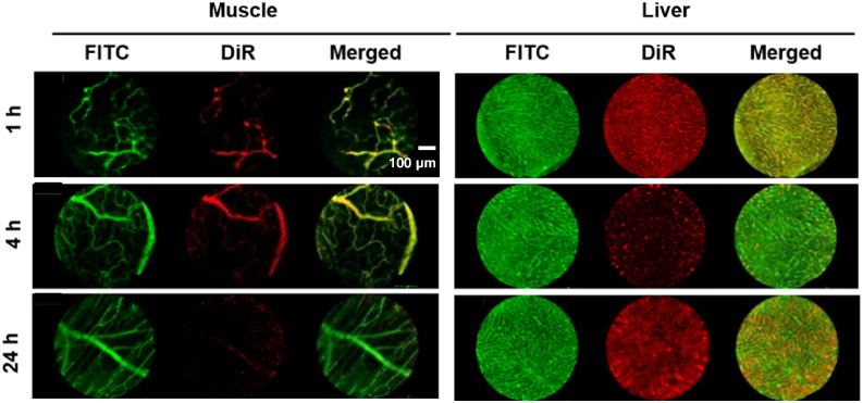
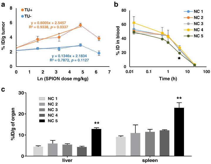
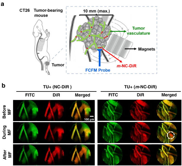
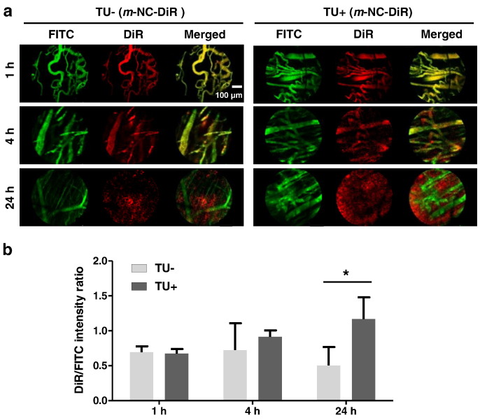
Magnetic drug targeting has been proposed as means of concentrating therapeutic agents at a target site and the success of this approach has been demonstrated in a number of studies. However, the behavior of magnetic carriers in blood vessels and tumor microcirculation still remains unclear. In this work, we utilized polymeric magnetic nanocapsules (m-NCs) for magnetic targeting in tumors and dynamically visualized them within blood vessels and tumor tissues before, during and after magnetic field exposure using fibered confocal fluorescence microscopy (FCFM). Our results suggested that the distribution of m-NCs within tumor vasculature changed dramatically, but in a reversible way, upon application and removal of a magnetic field. The m-NCs were concentrated and stayed as clusters near a blood vessel wall when tumors were exposed to a magnetic field but without rupturing the blood vessel. The obtained FCFM images provided in vivo in situ microvascular observations of m-NCs upon magnetic targeting with high spatial resolution but minimally invasive surgical procedures. This proof-of-concept descriptive study in mice is envisaged to track and quantify nanoparticles in vivo in a non-invasive manner at microscopic resolution.
磁靶向给药已被提议作为一种将治疗剂集中于靶位点的方法,并且该方法的成功已在多项研究中得到证实。然而,磁性载体在血管和肿瘤微循环中的行为仍不清楚。在这项工作中,我们利用聚合物磁性纳米胶囊(m-NCs)进行肿瘤的磁靶向,并使用纤维共聚焦荧光显微镜(FCFM)在磁场暴露之前、期间和之后动态观察它们在血管和肿瘤组织中的情况。我们的结果表明,在施加和去除磁场时,m-NCs在肿瘤脉管系统中的分布发生了显著但可逆的变化。当肿瘤暴露于磁场时,m-NCs聚集并以簇状停留在血管壁附近,但未使血管破裂。所获得的FCFM图像以高空间分辨率和微创外科手术方式提供了磁靶向时m-NCs在体内原位的微血管观察结果。设想这项在小鼠中的概念验证描述性研究能够以微观分辨率以非侵入性方式在体内追踪和定量纳米颗粒。