Bohîlțea R E, Cîrstoiu M M, Ciuvica A I, Munteanu O, Bodean O, Voicu D, Ionescu C A
"Carol Davila" University of Medicine and Pharmacy, Bucharest, Romania ; University Emergency Hospital Bucharest, Romania.
"Alfred Rusescu" Institute for Mother and Child Care, Bucharest, Romania.
J Med Life. 2016 Apr-Jun;9(2):126-9.
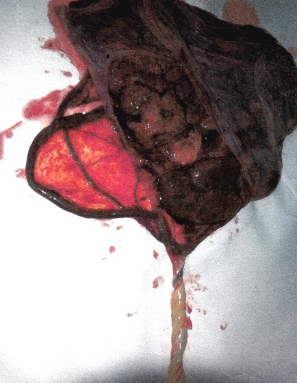
A velamentous umbilical cord is characterized by membranous umbilical vessels at the placental insertion site that are prone to compression and rupture, especially when they are located in the membranes covering the cervical os (vasa praevia). The velamentous insertion of the umbilical cord, with a reported incidence of 1% in singleton pregnancies and 15% in monochorionic twin gestations, has been associated with obstetric complications: fetal growth restriction, prematurity, congenital anomalies, low Apgar scores, fetal bleeding with acute fetal distress and placental retention. The pathogenesis is unknown, but the trophotropism theory is the most common and supported by the association of velamentous cord insertion and placenta praevia. The prevalence of vasa praevia is of approximately 1/ 2500 deliveries; the risk factors include the use of assisted reproductive technologies, low-lying placenta or placenta praevia, bilobed or succenturiate lobe placenta and multiple gestation. The diagnosis is rarely established before delivery and consequently the fetal mortality is extremely high. We report two cases of velamentous marginal umbilical cord insertion associated with vasa praevia (type 1 vasa praevia) and placenta praevia diagnosed during a routine mid-trimester fetal 2D ultrasound scan, color and power Doppler transvaginal ultrasound cervical assessment. The ultrasound examination revealed one umbilical vessel crossing the internal os of the cervix entering the placental margin and connecting to the subchorionic vasculature, remaining immobile when the uterus was shaken, the color Doppler imaging enhancing the identification of the vessel. The patients were admitted to the hospital in the third trimester and deliveries were planed and successfully performed at 38 weeks gestation, being confirmed by a macroscopic examination ultrasound diagnostic.
帆状脐带的特征是胎盘附着部位有膜状脐血管,这些血管容易受压和破裂,尤其是当它们位于覆盖宫颈内口的胎膜中时(前置血管)。脐带帆状附着在单胎妊娠中的发生率据报道为1%,在单绒毛膜双胎妊娠中为15%,与产科并发症有关:胎儿生长受限、早产、先天性异常、阿氏评分低、急性胎儿窘迫伴胎儿出血和胎盘滞留。其发病机制尚不清楚,但营养趋向性理论最为常见,且有帆状脐带附着与前置胎盘的关联作为支持。前置血管的发生率约为每2500例分娩中有1例;危险因素包括使用辅助生殖技术、低置胎盘或前置胎盘、双叶胎盘或副胎盘叶以及多胎妊娠。在分娩前很少能确诊,因此胎儿死亡率极高。我们报告两例帆状边缘性脐带附着合并前置血管(1型前置血管)和前置胎盘的病例,这是在孕中期常规胎儿二维超声扫描、经阴道彩色和能量多普勒超声宫颈评估时诊断出来的。超声检查显示一条脐血管穿过宫颈内口进入胎盘边缘并连接至绒毛膜下血管系统,当摇晃子宫时该血管保持不动,彩色多普勒成像增强了对该血管的识别。患者在孕晚期入院,计划在妊娠38周时分娩并顺利进行,经宏观检查证实超声诊断无误。