• 文献检索
  • 文档翻译
  • 深度研究
  • 学术资讯
  • Suppr Zotero 插件Zotero 插件
  • 邀请有礼
  • 套餐&价格
  • 历史记录
应用&插件
Suppr Zotero 插件Zotero 插件浏览器插件Mac 客户端Windows 客户端微信小程序
定价
高级版会员购买积分包购买API积分包
服务
文献检索文档翻译深度研究API 文档MCP 服务
关于我们
关于 Suppr公司介绍联系我们用户协议隐私条款
关注我们

Suppr 超能文献

核心技术专利:CN118964589B侵权必究
粤ICP备2023148730 号-1Suppr @ 2026

文献检索

告别复杂PubMed语法,用中文像聊天一样搜索,搜遍4000万医学文献。AI智能推荐,让科研检索更轻松。

立即免费搜索

文件翻译

保留排版,准确专业,支持PDF/Word/PPT等文件格式,支持 12+语言互译。

免费翻译文档

深度研究

AI帮你快速写综述,25分钟生成高质量综述,智能提取关键信息,辅助科研写作。

立即免费体验

利用磁共振成像分析来表征患有阻塞性睡眠呼吸暂停综合征的肥胖儿童的上气道结构。

MR Image Analytics to Characterize the Upper Airway Structure in Obese Children with Obstructive Sleep Apnea Syndrome.

作者信息

Tong Yubing, Udupa Jayaram K, Sin Sanghun, Liu Zhengbing, Wileyto E Paul, Torigian Drew A, Arens Raanan

机构信息

Medical Image Processing Group, Department of Radiology, University of Pennsylvania, Philadelphia, Pennsylvania, United States of America.

Division of Respiratory and Sleep Medicine, Children's Hospital at Montefiore, Bronx, New York, United States of America.

出版信息

PLoS One. 2016 Aug 3;11(8):e0159327. doi: 10.1371/journal.pone.0159327. eCollection 2016.

DOI:10.1371/journal.pone.0159327
PMID:27487240
原文链接:https://pmc.ncbi.nlm.nih.gov/articles/PMC4972248/
Abstract

PURPOSE

Quantitative image analysis in previous research in obstructive sleep apnea syndrome (OSAS) has focused on the upper airway or several objects in its immediate vicinity and measures of object size. In this paper, we take a more general approach of considering all major objects in the upper airway region and measures pertaining to their individual morphological properties, their tissue characteristics revealed by image intensities, and the 3D architecture of the object assembly. We propose a novel methodology to select a small set of salient features from this large collection of measures and demonstrate the ability of these features to discriminate with very high prediction accuracy between obese OSAS and obese non-OSAS groups.

MATERIALS AND METHODS

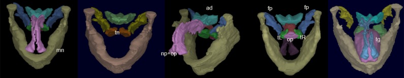
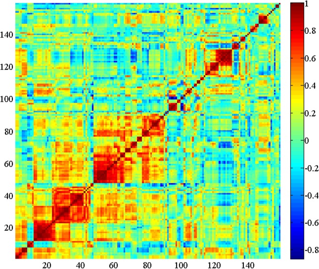
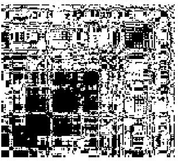
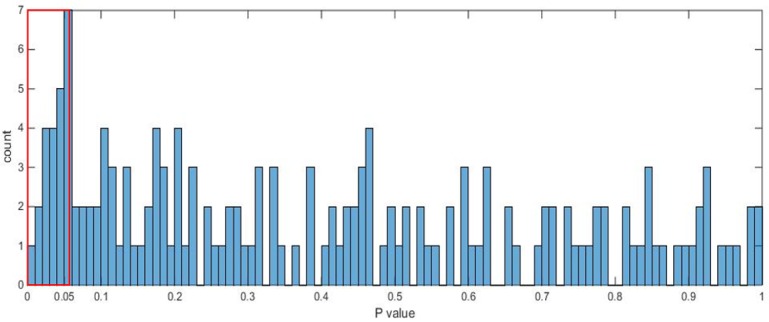
Thirty children were involved in this study with 15 in the obese OSAS group with an apnea-hypopnea index (AHI) = 14.4 ± 10.7) and 15 in the obese non-OSAS group with an AHI = 1.0 ± 1.0 (p<0.001). Subjects were between 8-17 years and underwent T1- and T2-weighted magnetic resonance imaging (MRI) of the upper airway during wakefulness. Fourteen objects in the vicinity of the upper airways were segmented in these images and a total of 159 measurements were derived from each subject image which included object size, surface area, volume, sphericity, standardized T2-weighted image intensity value, and inter-object distances. A small set of discriminating features was identified from this set in several steps. First, a subset of measures that have a low level of correlation among the measures was determined. A heat map visualization technique that allows grouping of parameters based on correlations among them was used for this purpose. Then, through T-tests, another subset of measures which are capable of separating the two groups was identified. The intersection of these subsets yielded the final feature set. The accuracy of these features to perform classification of unseen images into the two patient groups was tested by using logistic regression and multi-fold cross validation.

RESULTS

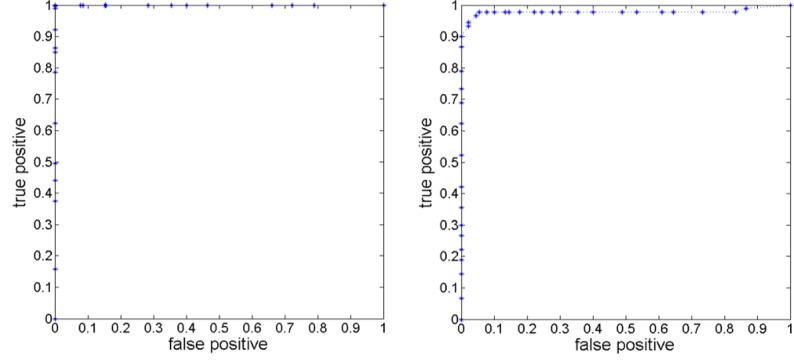
A set of 16 features identified with low inter-feature correlation (< 0.36) yielded a high classification accuracy of 96% with sensitivity and specificity of 97.8% and 94.4%, respectively. In addition to the previously observed increase in linear size, surface area, and volume of adenoid, tonsils, and fat pad in OSAS, the following new markers have been found. Standardized T2-weighted image intensities differed between the two groups for the entire neck body region, pharynx, and nasopharynx, possibly indicating changes in object tissue characteristics. Fat pad and oropharynx become less round or more complex in shape in OSAS. Fat pad and tongue move closer in OSAS, and so also oropharynx and tonsils and fat pad and tonsils. In contrast, fat pad and oropharynx move farther apart from the skin object.

CONCLUSIONS

The study has found several new anatomic bio-markers of OSAS. Changes in standardized T2-weighted image intensities in objects may imply that intrinsic tissue composition undergoes changes in OSAS. The results on inter-object distances imply that treatment methods should respect the relationships that exist among objects and not just their size. The proposed method of analysis may lead to an improved understanding of the mechanisms underlying OSAS.

摘要

目的

以往关于阻塞性睡眠呼吸暂停综合征(OSAS)的研究中的定量图像分析主要集中在上气道或其紧邻区域的几个对象以及对象大小的测量。在本文中,我们采用一种更通用的方法,考虑上气道区域中的所有主要对象以及与其各自形态特性、图像强度揭示的组织特征和对象集合的三维结构相关的测量。我们提出了一种新颖的方法,从这大量的测量中选择一小部分显著特征,并证明这些特征能够以非常高的预测准确率区分肥胖OSAS组和肥胖非OSAS组。

材料与方法

本研究纳入30名儿童,其中肥胖OSAS组15名,呼吸暂停低通气指数(AHI)为14.4±10.7,肥胖非OSAS组15名,AHI为1.0±1.0(p<0.001)。受试者年龄在8至17岁之间,清醒状态下接受上气道的T1加权和T2加权磁共振成像(MRI)检查。在这些图像中对上气道附近的14个对象进行分割,从每个受试者图像中总共获得159项测量值,包括对象大小、表面积、体积、球形度、标准化T2加权图像强度值以及对象间距离。通过几个步骤从这组测量值中确定一小部分有区分力的特征。首先,确定一组测量值之间相关性较低的子集。为此使用了一种热图可视化技术,该技术允许根据参数之间的相关性对参数进行分组。然后,通过T检验,确定另一组能够区分两组的测量值子集。这些子集的交集产生最终的特征集。通过逻辑回归和多折交叉验证测试这些特征将未见过的图像分类到两个患者组的准确性。

结果

一组16个特征,其特征间相关性较低(<0.36),分类准确率高达96%,敏感性和特异性分别为97.8%和94.4%。除了之前观察到的OSAS患者腺样体、扁桃体和脂肪垫的线性大小、表面积和体积增加外,还发现了以下新的标志物。两组在整个颈部身体区域、咽部和鼻咽部的标准化T2加权图像强度不同,这可能表明对象组织特征发生了变化。OSAS患者的脂肪垫和口咽形状变得不那么圆润或更复杂。OSAS患者中脂肪垫和舌靠近,口咽和扁桃体以及脂肪垫和扁桃体也靠近。相反,脂肪垫和口咽与皮肤对象的距离更远。

结论

该研究发现了几种新的OSAS解剖生物标志物。对象中标准化T2加权图像强度的变化可能意味着OSAS患者内在组织成分发生了变化。对象间距离的结果表明,治疗方法应考虑对象之间存在的关系,而不仅仅是它们的大小。所提出的分析方法可能有助于更好地理解OSAS的潜在机制。

https://cdn.ncbi.nlm.nih.gov/pmc/blobs/4691/4972248/aa6efb337a23/pone.0159327.g006.jpg
https://cdn.ncbi.nlm.nih.gov/pmc/blobs/4691/4972248/af425c62cf6c/pone.0159327.g001.jpg
https://cdn.ncbi.nlm.nih.gov/pmc/blobs/4691/4972248/f145af3efc28/pone.0159327.g002.jpg
https://cdn.ncbi.nlm.nih.gov/pmc/blobs/4691/4972248/d5bacbce7519/pone.0159327.g003.jpg
https://cdn.ncbi.nlm.nih.gov/pmc/blobs/4691/4972248/2cd485a4bd35/pone.0159327.g004.jpg
https://cdn.ncbi.nlm.nih.gov/pmc/blobs/4691/4972248/5b825421a0b0/pone.0159327.g005.jpg
https://cdn.ncbi.nlm.nih.gov/pmc/blobs/4691/4972248/aa6efb337a23/pone.0159327.g006.jpg

相似文献

1
MR Image Analytics to Characterize the Upper Airway Structure in Obese Children with Obstructive Sleep Apnea Syndrome.利用磁共振成像分析来表征患有阻塞性睡眠呼吸暂停综合征的肥胖儿童的上气道结构。
PLoS One. 2016 Aug 3;11(8):e0159327. doi: 10.1371/journal.pone.0159327. eCollection 2016.
2
Assessment of Upper-Airway Configuration in Obstructive Sleep Apnea Syndrome With Computed Tomography Imaging During Müller Maneuver.在Müller动作期间通过计算机断层扫描成像评估阻塞性睡眠呼吸暂停综合征的上气道形态
Respir Care. 2016 Dec;61(12):1651-1658. doi: 10.4187/respcare.04669. Epub 2016 Aug 16.
3
Understanding the anatomic basis for obstructive sleep apnea syndrome in adolescents.了解青少年阻塞性睡眠呼吸暂停综合征的解剖学基础。
Am J Respir Crit Care Med. 2015 Jun 1;191(11):1295-309. doi: 10.1164/rccm.201501-0169OC.
4
Magnetic resonance imaging of the upper airway structure of children with obstructive sleep apnea syndrome.阻塞性睡眠呼吸暂停综合征患儿上气道结构的磁共振成像
Am J Respir Crit Care Med. 2001 Aug 15;164(4):698-703. doi: 10.1164/ajrccm.164.4.2101127.
5
Minimally interactive segmentation of 4D dynamic upper airway MR images via fuzzy connectedness.通过模糊连接性对4D动态上呼吸道磁共振图像进行最小交互分割。
Med Phys. 2016 May;43(5):2323. doi: 10.1118/1.4945698.
6
Clinical, anthropometric and upper airway anatomic characteristics of obese patients with obstructive sleep apnea syndrome.阻塞性睡眠呼吸暂停综合征肥胖患者的临床、人体测量学及上气道解剖学特征
Respiration. 2007;74(5):517-24. doi: 10.1159/000097790. Epub 2006 Dec 5.
7
Adenotonsillectomy in obese children with obstructive sleep apnea syndrome: magnetic resonance imaging findings and considerations.肥胖儿童阻塞性睡眠呼吸暂停综合征的腺样体扁桃体切除术:磁共振成像的发现与考虑。
Sleep. 2013 Jun 1;36(6):841-7. doi: 10.5665/sleep.2708.
8
Upper airway structure and body fat composition in obese children with obstructive sleep apnea syndrome.肥胖儿童阻塞性睡眠呼吸暂停综合征的上气道结构和体脂成分。
Am J Respir Crit Care Med. 2011 Mar 15;183(6):782-7. doi: 10.1164/rccm.201008-1249OC. Epub 2010 Oct 8.
9
Evaluation of water content around airway in obstructive sleep apnea patients using peripharyngeal mucosal T2 magnetic resonance imaging.利用咽周黏膜T2磁共振成像评估阻塞性睡眠呼吸暂停患者气道周围的含水量
Clin Respir J. 2017 Nov;11(6):713-720. doi: 10.1111/crj.12405. Epub 2015 Oct 30.
10
Comparison of tongue volume/oral cavity volume ratio between obstructive sleep apnea syndrome patients and normal adults using magnetic resonance imaging.利用磁共振成像比较阻塞性睡眠呼吸暂停综合征患者与正常成年人的舌体积/口腔体积比。
J Med Dent Sci. 2006 Jun;53(2):119-26.

引用本文的文献

1
Mapping the Fat: How Childhood Obesity and Body Composition Shape Obstructive Sleep Apnoea.脂肪分布剖析:儿童肥胖与身体成分如何影响阻塞性睡眠呼吸暂停
Children (Basel). 2025 Jul 10;12(7):912. doi: 10.3390/children12070912.
2
Prediction of Radiation Therapy Induced Cardiovascular Toxicity from Pretreatment CT Images in Patients with Thoracic Malignancy via an Optimal Biomarker Approach.通过优化生物标志物方法,利用胸部恶性肿瘤患者治疗前CT图像预测放射治疗诱发的心血管毒性
Acad Radiol. 2025 Apr;32(4):1895-1905. doi: 10.1016/j.acra.2025.01.012. Epub 2025 Jan 26.
3
MRI-based radiomic features of the urinary bladder wall identify patients with moderate-to-severe international prostate symptom score.

本文引用的文献

1
The use of clinical parameters to predict obstructive sleep apnea syndrome severity in children: the Childhood Adenotonsillectomy (CHAT) study randomized clinical trial.利用临床参数预测儿童阻塞性睡眠呼吸暂停综合征的严重程度:儿童腺样体扁桃体切除术(CHAT)研究随机临床试验
JAMA Otolaryngol Head Neck Surg. 2015 Feb;141(2):130-6. doi: 10.1001/jamaoto.2014.3049.
2
Pediatric sleep-related breathing disorders: advances in imaging and computational modeling.小儿睡眠呼吸相关疾病:影像学与计算模型的进展
IEEE Pulse. 2014 Sep-Oct;5(5):33-9. doi: 10.1109/MPUL.2014.2339293.
3
Lateral cephalometric radiograph versus lateral nasopharyngeal radiograph for quantitative evaluation of nasopharyngeal airway space.
MRI 基于的膀胱壁放射组学特征可识别出具有中重度国际前列腺症状评分的患者。
World J Urol. 2024 Jun 13;42(1):375. doi: 10.1007/s00345-024-05081-3.
4
MRI-Based Radiomics Analysis of Levator Ani Muscle for Predicting Urine Incontinence after Robot-Assisted Radical Prostatectomy.基于MRI的肛提肌放射组学分析预测机器人辅助根治性前列腺切除术后尿失禁
Diagnostics (Basel). 2023 Sep 11;13(18):2913. doi: 10.3390/diagnostics13182913.
5
Optimal computed tomography-based biomarkers for prediction of incisional hernia formation.基于计算机断层扫描的最佳生物标志物预测切口疝形成。
Hernia. 2024 Feb;28(1):17-24. doi: 10.1007/s10029-023-02835-7. Epub 2023 Sep 7.
6
Neck fat and obstructive sleep apnea in obese adolescents.肥胖青少年的颈部脂肪与阻塞性睡眠呼吸暂停
Sleep. 2021 Nov 12;44(11). doi: 10.1093/sleep/zsab158.
7
Sleep-disordered breathing and comorbidities: role of the upper airway and craniofacial skeleton.睡眠呼吸障碍及其合并症:上呼吸道和颅面骨骼的作用
Nat Sci Sleep. 2020 Nov 9;12:907-936. doi: 10.2147/NSS.S146608. eCollection 2020.
8
Body region localization in whole-body low-dose CT images of PET/CT scans using virtual landmarks.使用虚拟标志物定位全身低剂量 CT 图像中的体部区域。
Med Phys. 2019 Mar;46(3):1286-1299. doi: 10.1002/mp.13376. Epub 2019 Jan 24.
9
Radiomics-guided therapy for bladder cancer: Using an optimal biomarker approach to determine extent of bladder cancer invasion from t2-weighted magnetic resonance images.基于影像组学的膀胱癌治疗:采用优化生物标志物方法从T2加权磁共振图像确定膀胱癌浸润范围
Adv Radiat Oncol. 2018 May 8;3(3):331-338. doi: 10.1016/j.adro.2018.04.011. eCollection 2018 Jul-Sep.
用于鼻咽气道空间定量评估的头颅侧位X线片与鼻咽侧位X线片对比
Dental Press J Orthod. 2014 Jul-Aug;19(4):89-93. doi: 10.1590/2176-9451.19.4.089-093.oar.
4
A texture analysis method for MR images of airway dilator muscles: a feasibility study.气道扩张肌磁共振成像的纹理分析方法:一项可行性研究。
Dentomaxillofac Radiol. 2014;43(5):20130403. doi: 10.1259/dmfr.20130403. Epub 2014 Apr 29.
5
Quantitative upper airway endoscopy with swept-source anatomical optical coherence tomography.采用扫频源解剖光学相干断层扫描技术的定量上气道内窥镜检查
Biomed Opt Express. 2014 Feb 19;5(3):788-99. doi: 10.1364/BOE.5.000788. eCollection 2014 Mar 1.
6
Prevalence of childhood and adult obesity in the United States, 2011-2012.美国儿童和成人肥胖率,2011-2012 年。
JAMA. 2014 Feb 26;311(8):806-14. doi: 10.1001/jama.2014.732.
7
Predictors of obstructive sleep apnea severity in adenotonsillectomy candidates.腺样体扁桃体切除术候选患者阻塞性睡眠呼吸暂停严重程度的预测因素。
Sleep. 2014 Feb 1;37(2):261-9. doi: 10.5665/sleep.3394.
8
Adenotonsillectomy in obese children with obstructive sleep apnea syndrome: magnetic resonance imaging findings and considerations.肥胖儿童阻塞性睡眠呼吸暂停综合征的腺样体扁桃体切除术:磁共振成像的发现与考虑。
Sleep. 2013 Jun 1;36(6):841-7. doi: 10.5665/sleep.2708.
9
Physiological effects of obstructive sleep apnea syndrome in childhood.儿童阻塞性睡眠呼吸暂停综合征的生理影响。
Respir Physiol Neurobiol. 2013 Sep 15;188(3):370-82. doi: 10.1016/j.resp.2013.05.006. Epub 2013 May 23.
10
Effective three-dimensional evaluation analysis of upper airway form during oral appliance therapy in patients with obstructive sleep apnoea.口腔矫治器治疗阻塞性睡眠呼吸暂停患者上气道形态的有效三维评价分析。
J Oral Rehabil. 2013 Aug;40(8):582-9. doi: 10.1111/joor.12059. Epub 2013 May 21.