Department of Bioengineering, School of Engineering and Applied Sciences, University of Pennsylvania. Philadelphia, Pennsylvania, United States.
Department of Neurosurgery, Perelman School of Medicine, University of Pennsylvania, Philadelphia, Pennsylvania, United States.
Sci Rep. 2016 Aug 8;6:31215. doi: 10.1038/srep31215.
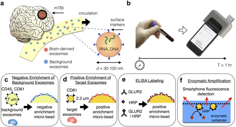
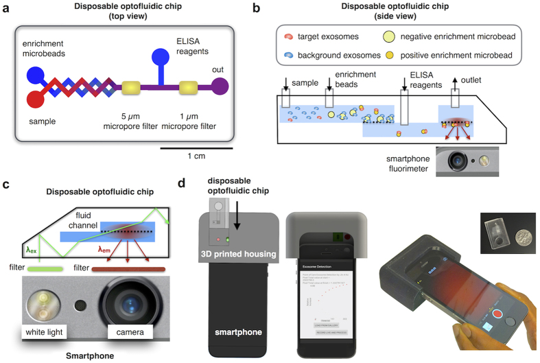
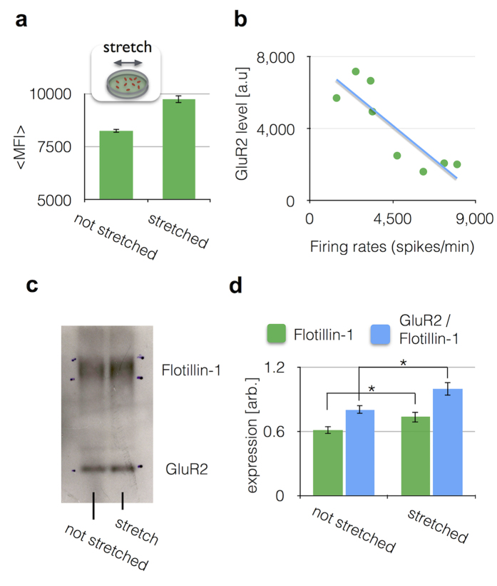
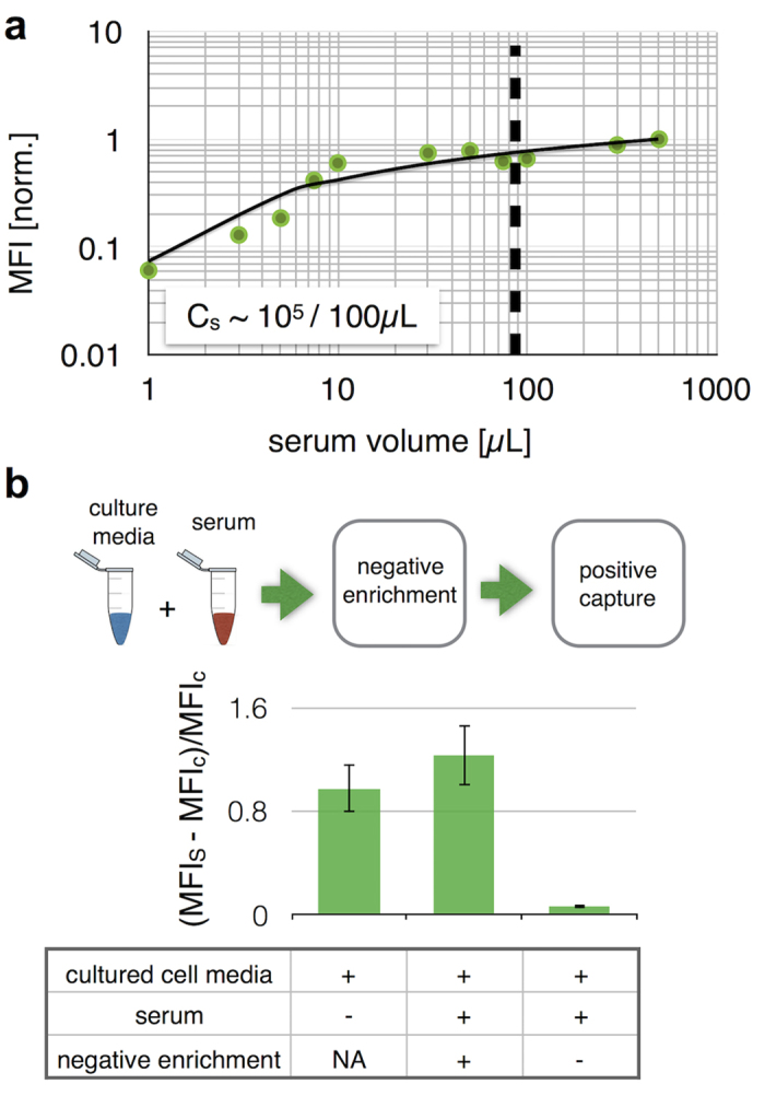
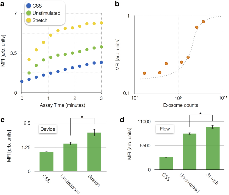
A major impediment to improving the treatment of concussion is our current inability to identify patients that will experience persistent problems after the injury. Recently, brain-derived exosomes, which cross the blood-brain barrier and circulate following injury, have shown great potential as a noninvasive biomarker of brain recovery. However, clinical use of exosomes has been constrained by their small size (30-100 nm) and the extensive sample preparation (>24 hr) needed for traditional exosome measurements. To address these challenges, we developed a smartphone-enabled optofluidic platform to measure brain-derived exosomes. Sample-to-answer on our chip is 1 hour, 10x faster than conventional techniques. The key innovation is an optofluidic device that can detect enzyme amplified exosome biomarkers, and is read out using a smartphone camera. Using this approach, we detected and profiled GluR2+ exosomes in the post-injury state using both in vitro and murine models of concussion.
改善脑震荡治疗的主要障碍是我们目前无法识别在受伤后会出现持续问题的患者。最近,脑源性外泌体在损伤后穿过血脑屏障并循环,作为脑恢复的非侵入性生物标志物显示出巨大的潜力。然而,外泌体的临床应用受到其小尺寸(30-100nm)和传统外泌体测量所需的广泛样本制备(>24 小时)的限制。为了解决这些挑战,我们开发了一种智能手机启用的光流控平台来测量脑源性外泌体。我们的芯片上实现了 1 小时的样本到答案,比传统技术快 10 倍。关键的创新是一种光流控装置,可以检测酶放大的外泌体生物标志物,并使用智能手机摄像头进行读取。使用这种方法,我们使用体外和小鼠脑震荡模型在受伤后状态下检测和分析了 GluR2+外泌体。