Shalaby Mona Fattouh Mohamed, El-Din Asmaa Nasr, El-Hamd Mohammed Abu
Department of Medical Microbiology and Immunology, Faculty of Medicine, Sohag University, Egypt.
Department of Dermatology, Venereology and Andrology, Faculty of Medicine, Sohag University, Egypt.
Electron Physician. 2016 Jun 25;8(6):2557-67. doi: 10.19082/2557. eCollection 2016 Jun.
The objective of this study was to isolate, identify, and explore the in-vitro antifungal susceptibility pattern of dermatophytes isolated from clinically suspected cases of dermatophytosis (tinea infections) attending the Dermatology Outpatient Clinic.
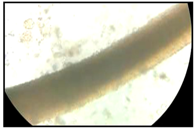
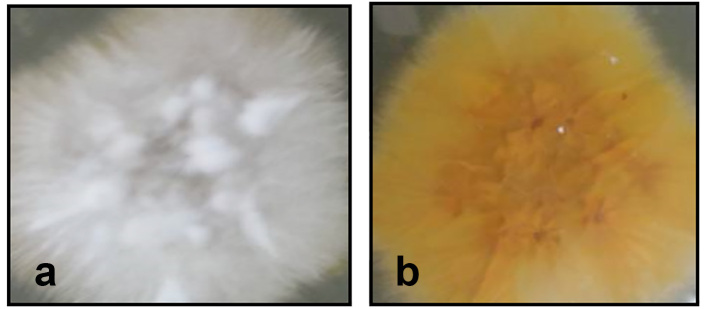
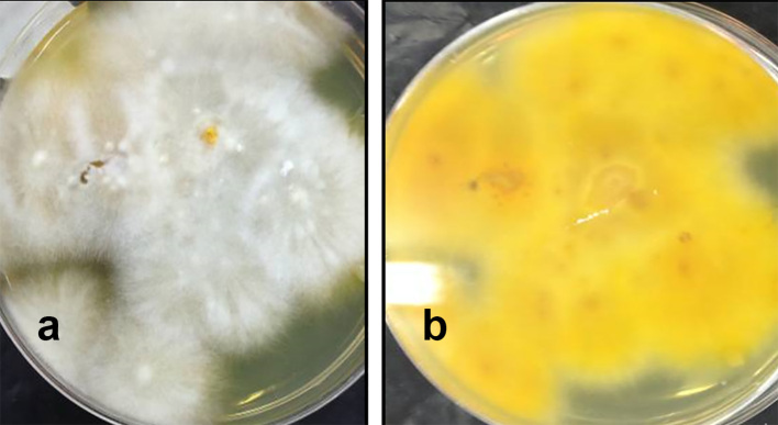
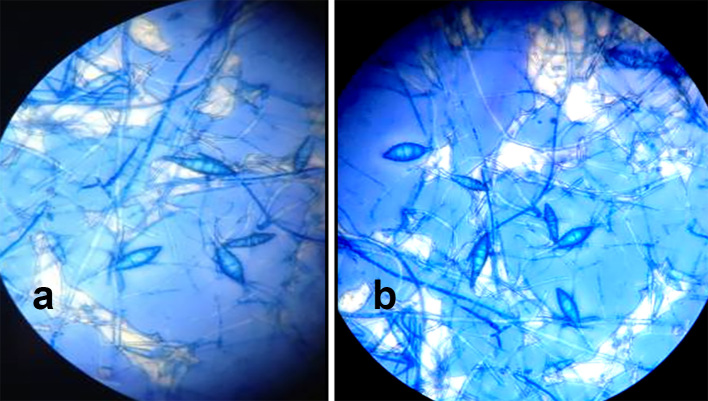
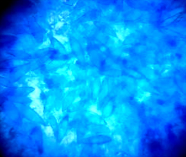
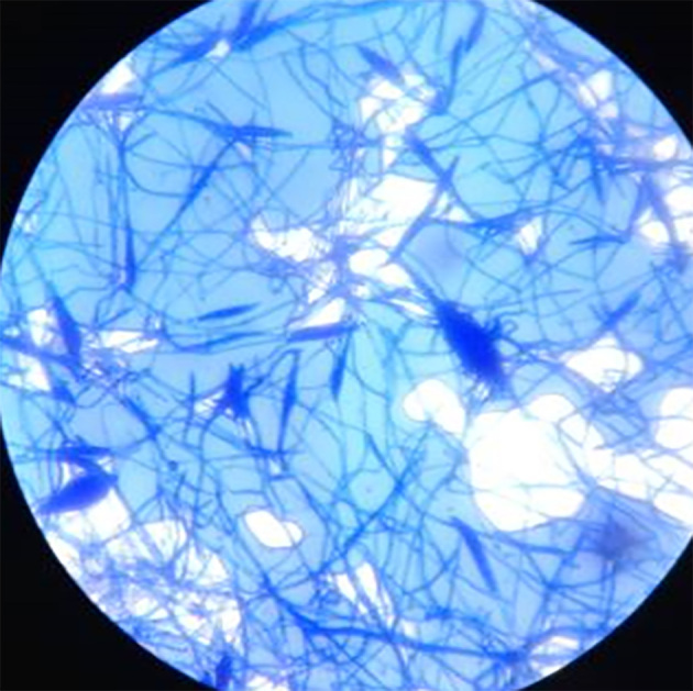
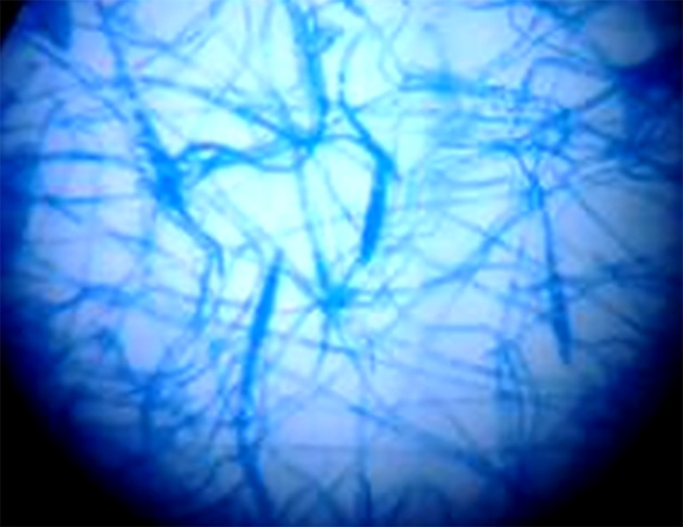
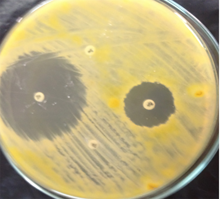
This study was conducted at Sohag University Hospital from December 2014 to December 2015. Clinical samples (e.g., skin scrapings and hair stumps) were collected under aseptic precautions. The identification of dermatophytes was performed through microscopic examination using 10% potassium hydroxide (KOH) with 40% dimethyl sulphoxide (DMSO) mounts and culture on Sabouraud dextrose agar (SDA) and on Dermasel agar base media, both supplemented with chloramphenicol and cycloheximide. All dermatophytes isolates were subjected to antifungal susceptibility testing using the agar-based disk diffusion (ABDD) method against Clotrimazole, Miconazole, Fluconazole, and Griseofulvin. Data were analyzed via SPSS 16, using Chi square and a screening test (cross-tabulation method).
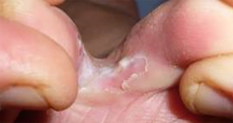
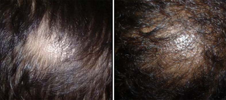
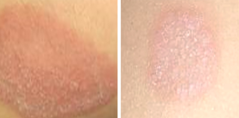
A total of 110 patients of dermatophytosis were studied. The patients were clinically diagnosed and mycologically confirmed as having tinea capitis (49), tinea corporis (30), tinea pedis (16), tinea cruris (9), or tinea barbae (6). The dermatophytes isolates belonged to 4 species: Microsporum canis 58 (52.7%), Microsporum gypseum 23 (20.9%), Trichophyton mentagrophytes 18 (16.4%), and Microsporum audouinii 11 (10%). The most effective antifungal drugs tested were Clotrimazole, followed by Miconazole (95.5% and 84.5% of isolates were susceptible, respectively).
Every patient with a tinea infection should be properly studied for a mycological examination and should be treated accordingly. Dermasel agar is more useful as an identification medium in the isolation of dermatophytes. The ABDD method appears to be a simple, cost-effective, and promising method for the evaluation of antifungal susceptibility of dermatophytes.
本研究的目的是从皮肤科门诊临床疑似皮肤癣菌病(癣感染)病例中分离、鉴定皮肤癣菌,并探讨其体外抗真菌药敏模式。
本研究于2014年12月至2015年12月在索哈杰大学医院进行。在无菌预防措施下收集临床样本(如皮肤刮屑和毛发残根)。通过使用10%氢氧化钾(KOH)加40%二甲基亚砜(DMSO)制片进行显微镜检查,并在添加了氯霉素和放线菌酮的沙氏葡萄糖琼脂(SDA)和皮肤癣菌琼脂基础培养基上培养来鉴定皮肤癣菌。所有皮肤癣菌分离株均采用基于琼脂的纸片扩散法(ABDD)对克霉唑、咪康唑、氟康唑和灰黄霉素进行抗真菌药敏试验。数据通过SPSS 16使用卡方检验和筛选试验(交叉表法)进行分析。
共研究了110例皮肤癣菌病患者。患者经临床诊断和真菌学确诊为头癣(49例)、体癣(30例)、足癣(16例)、股癣(9例)或须癣(6例)。皮肤癣菌分离株属于4个种:犬小孢子菌58株(52.7%)、石膏样小孢子菌23株(20.9%)、须癣毛癣菌18株(16.4%)和奥杜盎小孢子菌11株(10%)。所测试的最有效的抗真菌药物是克霉唑,其次是咪康唑(分别有95.5%和84.5%的分离株敏感)。
每例癣感染患者都应进行适当的真菌学检查并相应治疗。皮肤癣菌琼脂在分离皮肤癣菌时作为鉴定培养基更有用。ABDD法似乎是一种简单、经济有效且有前景的评估皮肤癣菌抗真菌药敏的方法。