Papp Stefanie, Moderzynski Kristin, Rauch Jessica, Heine Liza, Kuehl Svenja, Richardt Ulricke, Mueller Heidelinde, Fleischer Bernhard, Osterloh Anke
Department of Immunology, Bernhard Nocht Institute for Tropical Medicine, Hamburg, Germany.
Institute for Immunology, University Medical Center Hamburg-Eppendorf, Hamburg, Germany.
PLoS Negl Trop Dis. 2016 Aug 22;10(8):e0004935. doi: 10.1371/journal.pntd.0004935. eCollection 2016 Aug.
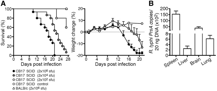
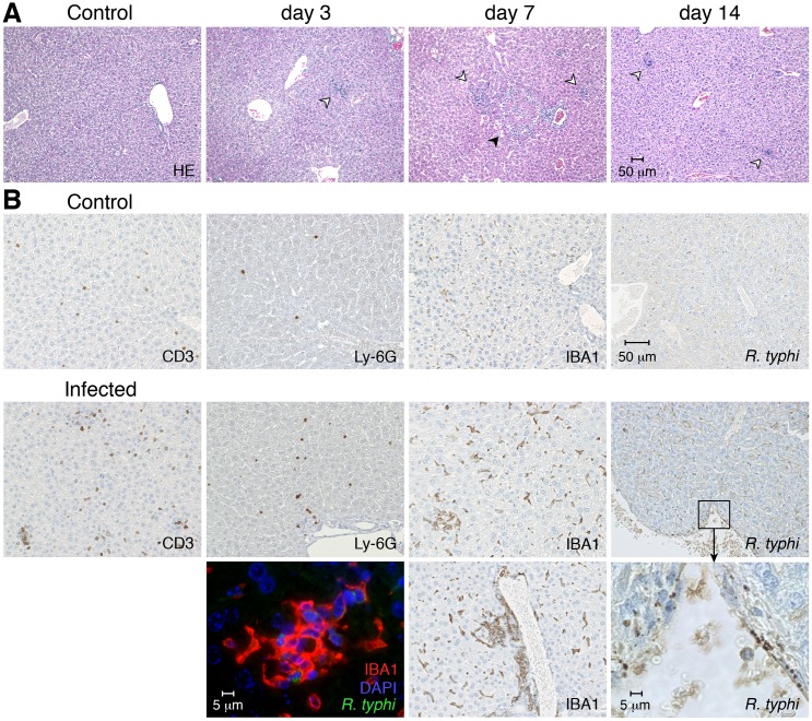
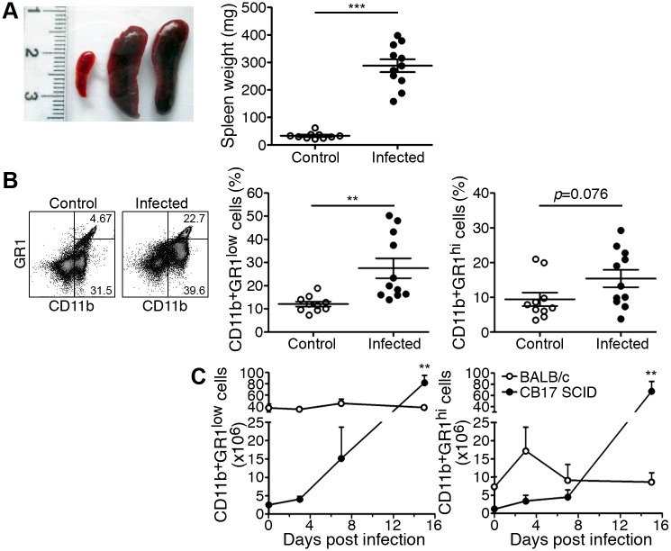
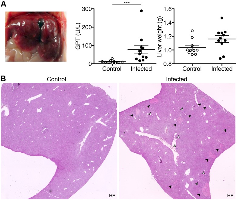
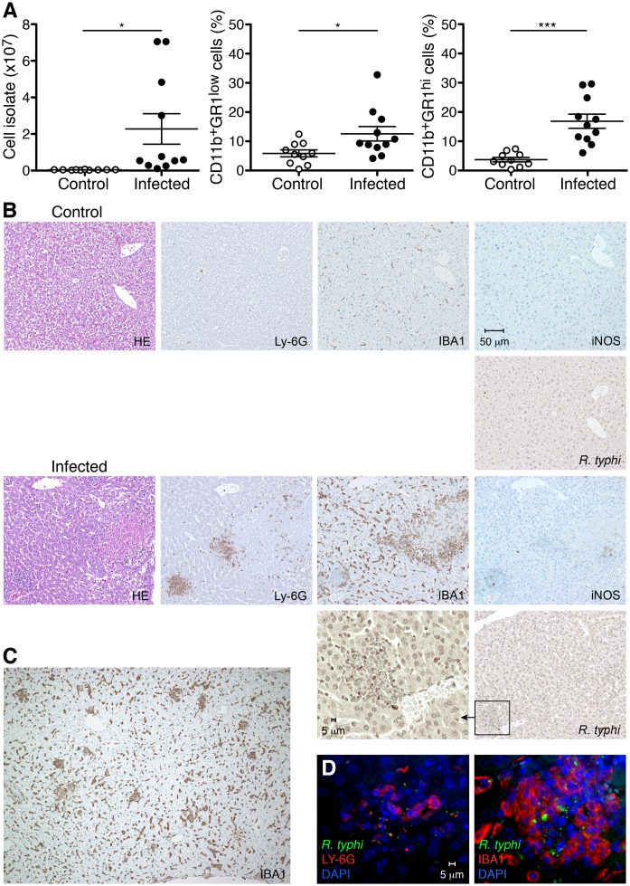
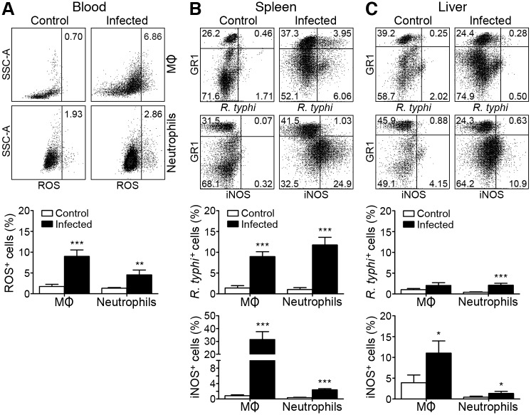
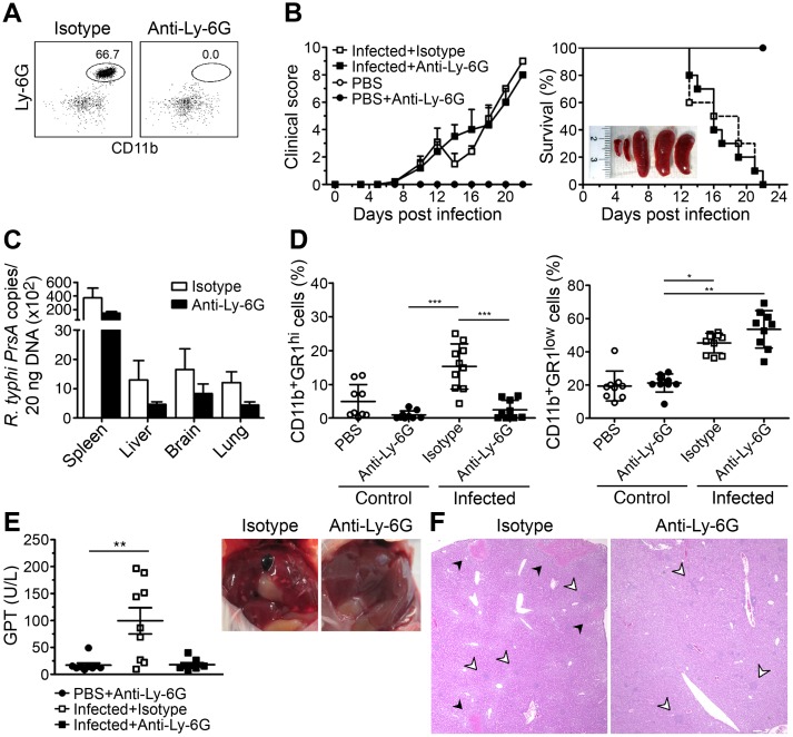
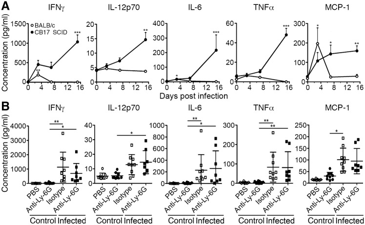
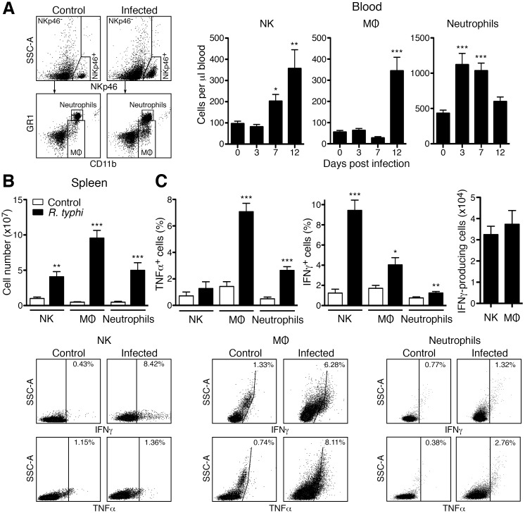
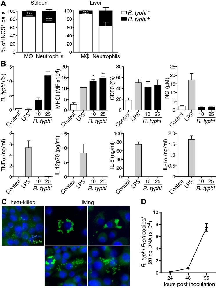
Rickettsia (R.) typhi is the causative agent of endemic typhus, an emerging febrile disease that is associated with complications such as pneumonia, encephalitis and liver dysfunction. To elucidate how innate immune mechanisms contribute to defense and pathology we here analyzed R. typhi infection of CB17 SCID mice that are congenic to BALB/c mice but lack adaptive immunity. CB17 SCID mice succumbed to R. typhi infection within 21 days and showed high bacterial load in spleen, brain, lung, and liver. Most evident pathological changes in R. typhi-infected CB17 SCID mice were massive liver necrosis and splenomegaly due to the disproportionate accumulation of neutrophils and macrophages (MΦ). Both neutrophils and MΦ infiltrated the liver and harbored R. typhi. Both cell populations expressed iNOS and produced reactive oxygen species (ROS) and, thus, exhibited an inflammatory and bactericidal phenotype. Surprisingly, depletion of neutrophils completely prevented liver necrosis but neither altered bacterial load nor protected CB17 SCID mice from death. Furthermore, the absence of neutrophils had no impact on the overwhelming systemic inflammatory response in these mice. This response was predominantly driven by activated MΦ and NK cells both of which expressed IFNγ and is considered as the reason of death. Finally, we observed that iNOS expression by MΦ and neutrophils did not correlate with R. typhi uptake in vivo. Moreover, we demonstrate that MΦ hardly respond to R. typhi in vitro. These findings indicate that R. typhi enters MΦ and also neutrophils unrecognized and that activation of these cells is mediated by other mechanisms in the context of tissue damage in vivo.
鼠型斑疹伤寒立克次体是地方性斑疹伤寒的病原体,这是一种新出现的发热性疾病,与肺炎、脑炎和肝功能障碍等并发症相关。为了阐明固有免疫机制如何促进防御和病理过程,我们在此分析了CB17 SCID小鼠的鼠型斑疹伤寒立克次体感染情况,这些小鼠与BALB/c小鼠同源,但缺乏适应性免疫。CB17 SCID小鼠在21天内死于鼠型斑疹伤寒立克次体感染,并在脾脏、大脑、肺和肝脏中显示出高细菌载量。鼠型斑疹伤寒立克次体感染的CB17 SCID小鼠最明显的病理变化是由于中性粒细胞和巨噬细胞(MΦ)不成比例的积聚导致的大量肝坏死和脾肿大。中性粒细胞和MΦ都浸润肝脏并携带鼠型斑疹伤寒立克次体。这两种细胞群体均表达诱导型一氧化氮合酶(iNOS)并产生活性氧(ROS),因此表现出炎症和杀菌表型。令人惊讶的是,中性粒细胞的耗竭完全预防了肝坏死,但既未改变细菌载量,也未保护CB17 SCID小鼠免于死亡。此外,中性粒细胞的缺失对这些小鼠中压倒性的全身炎症反应没有影响。这种反应主要由活化的MΦ和自然杀伤(NK)细胞驱动,这两种细胞均表达γ干扰素(IFNγ),并被认为是死亡原因。最后,我们观察到MΦ和中性粒细胞的iNOS表达与体内鼠型斑疹伤寒立克次体摄取无关。此外,我们证明MΦ在体外对鼠型斑疹伤寒立克次体几乎没有反应。这些发现表明,鼠型斑疹伤寒立克次体进入MΦ以及中性粒细胞未被识别,并且这些细胞的激活是在体内组织损伤的背景下由其他机制介导的。