• 文献检索
  • 文档翻译
  • 深度研究
  • 学术资讯
  • Suppr Zotero 插件Zotero 插件
  • 邀请有礼
  • 套餐&价格
  • 历史记录
应用&插件
Suppr Zotero 插件Zotero 插件浏览器插件Mac 客户端Windows 客户端微信小程序
定价
高级版会员购买积分包购买API积分包
服务
文献检索文档翻译深度研究API 文档MCP 服务
关于我们
关于 Suppr公司介绍联系我们用户协议隐私条款
关注我们

Suppr 超能文献

核心技术专利:CN118964589B侵权必究
粤ICP备2023148730 号-1Suppr @ 2026

文献检索

告别复杂PubMed语法,用中文像聊天一样搜索,搜遍4000万医学文献。AI智能推荐,让科研检索更轻松。

立即免费搜索

文件翻译

保留排版,准确专业,支持PDF/Word/PPT等文件格式,支持 12+语言互译。

免费翻译文档

深度研究

AI帮你快速写综述,25分钟生成高质量综述,智能提取关键信息,辅助科研写作。

立即免费体验

小鼠光学相干断层扫描血管造影:与共聚焦扫描激光显微镜和荧光素血管造影的比较。

Optical Coherence Tomography Angiography in Mice: Comparison with Confocal Scanning Laser Microscopy and Fluorescein Angiography.

作者信息

Giannakaki-Zimmermann Helena, Kokona Despina, Wolf Sebastian, Ebneter Andreas, Zinkernagel Martin S

机构信息

Department of Ophthalmology and Department of Clinical Research Inselspital, Bern University Hospital, and University of Bern, Switzerland.

出版信息

Transl Vis Sci Technol. 2016 Aug 18;5(4):11. doi: 10.1167/tvst.5.4.11. eCollection 2016 Aug.

DOI:10.1167/tvst.5.4.11
PMID:27570710
原文链接:https://pmc.ncbi.nlm.nih.gov/articles/PMC4997887/
Abstract

PURPOSE

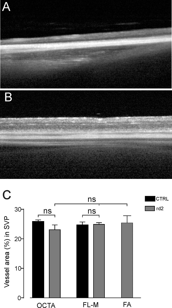
Optical coherence tomography angiography (OCT-A) allows noninvasive visualization of retinal vessels in vivo. OCT-A was used to characterize the vascular network of the mouse retina and was compared with fluorescein angiography (FA) and histology.

METHODS

In the present study, OCT-A based on a Heidelberg Engineering Spectralis system was used to investigate the vascular network in mice. Data was compared with FA and confocal microscopy of flat-mount histology stained with isolectin IB4. For quantitative analysis the National Cancer Institute's AngioTool software was used. Vessel density, the number of vessel junctions, and endpoints were measured and compared between the imaging modalities.

RESULTS

The configuration of the superficial capillary network was comparable with OCT-A and flat-mount histology in BALBc mice. However, vessel density and the number of vessel junctions per region of interest ( = 0.0161 and = 0.0015, respectively) in the deep vascular network of BALBc mice measured by OCT-A was significantly higher than with flat-mount histology. In C3A.Cg mice, where the deep capillary plexus is absent, analysis of the superficial network provided similar results for all three imaging modalities.

CONCLUSION

OCT-A is a helpful imaging tool for noninvasive, in vivo imaging of the vascular plexus in mice. It may offer advantages over FA and confocal microscopy especially for imaging the deep vascular plexus.

TRANSLATIONAL RELEVANCE

The present study shows that OCT-A can be employed for small animal imaging to assess the vascular network and offers advantages over flat-mount histology and FA.

摘要

目的

光学相干断层扫描血管造影术(OCT-A)可在体内对视网膜血管进行无创可视化。本研究使用OCT-A对小鼠视网膜血管网络进行特征分析,并与荧光素血管造影术(FA)和组织学检查进行比较。

方法

在本研究中,基于海德堡工程公司Spectralis系统的OCT-A被用于研究小鼠的血管网络。将数据与FA以及用异凝集素IB4染色的平铺组织学共聚焦显微镜检查结果进行比较。使用美国国立癌症研究所的AngioTool软件进行定量分析。测量并比较了不同成像方式下的血管密度、血管交叉点数量和端点数量。

结果

BALBc小鼠的浅表毛细血管网络结构在OCT-A和平铺组织学检查中具有可比性。然而,通过OCT-A测量的BALBc小鼠深层血管网络中每个感兴趣区域的血管密度和血管交叉点数量(分别为=0.0161和=0.0015)显著高于平铺组织学检查结果。在缺乏深层毛细血管丛的C3A.Cg小鼠中,对浅表网络的分析在所有三种成像方式下均得到了相似的结果。

结论

OCT-A是一种用于小鼠血管丛无创体内成像的有用成像工具。它可能比FA和共聚焦显微镜具有优势,特别是在对深层血管丛成像方面。

转化相关性

本研究表明,OCT-A可用于小动物成像以评估血管网络,并且比平铺组织学检查和FA具有优势。

https://cdn.ncbi.nlm.nih.gov/pmc/blobs/7c42/4997887/cc15d069870b/i2164-2591-5-4-11-f04.jpg

相似文献

1
Optical Coherence Tomography Angiography in Mice: Comparison with Confocal Scanning Laser Microscopy and Fluorescein Angiography.小鼠光学相干断层扫描血管造影:与共聚焦扫描激光显微镜和荧光素血管造影的比较。
Transl Vis Sci Technol. 2016 Aug 18;5(4):11. doi: 10.1167/tvst.5.4.11. eCollection 2016 Aug.
2
Optical coherence tomography angiography (OCT-A) in an animal model of laser-induced choroidal neovascularization.光学相干断层扫描血管造影(OCT-A)在激光诱导脉络膜新生血管动物模型中的应用。
Exp Eye Res. 2019 Jul;184:162-171. doi: 10.1016/j.exer.2019.04.002. Epub 2019 Apr 16.
3
Angiography reveals novel features of the retinal vasculature in healthy and diabetic mice.血管造影揭示了健康小鼠和糖尿病小鼠视网膜血管系统的新特征。
Exp Eye Res. 2015 Sep;138:6-21. doi: 10.1016/j.exer.2015.06.023. Epub 2015 Jun 26.
4
Colocalization of optical coherence tomography angiography with histology in the mouse retina.光学相干断层扫描血管造影与组织学在小鼠视网膜中的共定位。
Microvasc Res. 2020 Nov;132:104055. doi: 10.1016/j.mvr.2020.104055. Epub 2020 Aug 8.
5
OCT angiography in the mouse: A novel evaluation method for vascular pathologies of the mouse retina.小鼠的光学相干断层扫描血管造影:一种用于评估小鼠视网膜血管病变的新方法。
Exp Eye Res. 2016 Apr;145:417-423. doi: 10.1016/j.exer.2016.02.012. Epub 2016 Mar 2.
6
Optical Coherence Tomography Angiography in Retinal Vein Occlusion: Evaluation of Superficial and Deep Capillary Plexa.视网膜静脉阻塞中的光学相干断层扫描血管造影:浅层和深层毛细血管丛的评估
Am J Ophthalmol. 2016 Jan;161:160-71.e1-2. doi: 10.1016/j.ajo.2015.10.008. Epub 2015 Oct 23.
7
In vivo optical imaging of human retinal capillary networks using speckle variance optical coherence tomography with quantitative clinico-histological correlation.使用散斑方差光学相干断层扫描对人视网膜毛细血管网络进行体内光学成像,并进行定量临床组织学相关性分析。
Microvasc Res. 2015 Jul;100:32-9. doi: 10.1016/j.mvr.2015.04.006. Epub 2015 Apr 24.
8
Retinal vascular layers imaged by fluorescein angiography and optical coherence tomography angiography.荧光素血管造影和光相干断层扫描血管造影成像的视网膜血管层。
JAMA Ophthalmol. 2015 Jan;133(1):45-50. doi: 10.1001/jamaophthalmol.2014.3616.
9
OCT Angiography: Measurement of Retinal Macular Microvasculature with Spectralis II OCT Angiography - Reliability and Reproducibility.OCT 血管造影:应用 Spectralis II OCT 血管造影测量视网膜黄斑微循环 - 可靠性和可重复性。
Ophthalmologica. 2020;243(1):75-84. doi: 10.1159/000502458. Epub 2019 Sep 11.
10
Comparisons Between Histology and Optical Coherence Tomography Angiography of the Periarterial Capillary-Free Zone.periarterial capillary-free zone 组织学与光相干断层扫描血管造影比较
Am J Ophthalmol. 2018 May;189:55-64. doi: 10.1016/j.ajo.2018.02.007. Epub 2018 Feb 19.

引用本文的文献

1
Effects of Treadmill Exercise on Retinal Vascular Morphology, Function, and Circulating Immune Factors in a Mouse Model of Retinal Degeneration.跑步机运动对视网膜变性小鼠模型视网膜血管形态、功能及循环免疫因子的影响
Invest Ophthalmol Vis Sci. 2025 Aug 1;66(11):65. doi: 10.1167/iovs.66.11.65.
2
Characterization of the Retinal Circulation of the Mouse.小鼠视网膜循环的特征描述
Invest Ophthalmol Vis Sci. 2024 Dec 2;65(14):3. doi: 10.1167/iovs.65.14.3.
3
OCT and ERG Techniques in High-Throughput Phenotyping of Mouse Vision.OCT 和 ERG 技术在小鼠视觉高通量表型分析中的应用。

本文引用的文献

1
Longitudinal and Cross-Sectional Analyses of Age Effects on Retinal Nerve Fiber Layer and Ganglion Cell Complex Thickness by Fourier-Domain OCT.傅里叶域光学相干断层扫描对年龄对视网膜神经纤维层和神经节细胞复合体厚度影响的纵向和横断面分析
Transl Vis Sci Technol. 2016 Mar 4;5(2):1. doi: 10.1167/tvst.5.2.1. eCollection 2016 Mar.
2
OCT angiography in the mouse: A novel evaluation method for vascular pathologies of the mouse retina.小鼠的光学相干断层扫描血管造影:一种用于评估小鼠视网膜血管病变的新方法。
Exp Eye Res. 2016 Apr;145:417-423. doi: 10.1016/j.exer.2016.02.012. Epub 2016 Mar 2.
3
RETINAL LAYER RESPONSE TO RANIBIZUMAB DURING TREATMENT OF DIABETIC MACULAR EDEMA: Thinner is Not Always Better.
Genes (Basel). 2023 Jan 22;14(2):294. doi: 10.3390/genes14020294.
4
Endothelial Toll-like receptor 4 is required for microglia activation in the murine retina after systemic lipopolysaccharide exposure.内皮 Toll 样受体 4 是系统性脂多糖暴露后小鼠视网膜小胶质细胞激活所必需的。
J Neuroinflammation. 2023 Feb 4;20(1):25. doi: 10.1186/s12974-023-02712-1.
5
Methods to measure blood flow and vascular reactivity in the retina.测量视网膜血流和血管反应性的方法。
Front Med (Lausanne). 2023 Jan 12;9:1069449. doi: 10.3389/fmed.2022.1069449. eCollection 2022.
6
Selective vulnerability of the intermediate retinal capillary plexus precedes retinal ganglion cell loss in ocular hypertension.在高眼压症中,视网膜中间毛细血管丛的选择性易损性先于视网膜神经节细胞丢失。
Front Cell Neurosci. 2022 Dec 5;16:1073786. doi: 10.3389/fncel.2022.1073786. eCollection 2022.
7
Insights into Metabolic Activity and Structure of the Retina through Multiphoton Fluorescence Lifetime Imaging Microscopy in Mice.通过多光子荧光寿命成像显微镜观察小鼠视网膜的代谢活性和结构。
Cells. 2022 Jul 22;11(15):2265. doi: 10.3390/cells11152265.
8
Morphological and vascular characteristics of the optic nerve head of normal guinea pigs.正常豚鼠视神经乳头的形态和血管特征。
Sci Rep. 2022 Jan 18;12(1):873. doi: 10.1038/s41598-022-04911-x.
9
Characterization of the Canine Retinal Vasculature With Optical Coherence Tomography Angiography: Comparisons With Histology and Fluorescein Angiography.光学相干断层扫描血管造影对犬视网膜血管系统的特征描述:与组织学和荧光素血管造影的比较
Front Neuroanat. 2021 Dec 13;15:785249. doi: 10.3389/fnana.2021.785249. eCollection 2021.
10
Morphometric and Microstructural Changes During Murine Retinal Development Characterized Using In Vivo Optical Coherence Tomography.利用活体光学相干断层扫描技术对小鼠视网膜发育过程中的形态和微观结构变化进行研究。
Invest Ophthalmol Vis Sci. 2021 Oct 4;62(13):20. doi: 10.1167/iovs.62.13.20.
糖尿病性黄斑水肿治疗期间视网膜各层对雷珠单抗的反应:并非越薄越好。
Retina. 2016 Jul;36(7):1314-23. doi: 10.1097/IAE.0000000000000923.
4
Volume-Rendered Optical Coherence Tomography of Diabetic Retinopathy Pilot Study.糖尿病视网膜病变的容积渲染光学相干断层扫描试点研究
Am J Ophthalmol. 2015 Dec;160(6):1200-10. doi: 10.1016/j.ajo.2015.09.010. Epub 2015 Sep 15.
5
Quantitative Analysis of Mouse Retinal Layers Using Automated Segmentation of Spectral Domain Optical Coherence Tomography Images.使用光谱域光学相干断层扫描图像的自动分割对小鼠视网膜层进行定量分析
Transl Vis Sci Technol. 2015 Aug 25;4(4):9. doi: 10.1167/tvst.4.4.9. eCollection 2015 Aug.
6
The Role of LOX and LOXL2 in the Pathogenesis of an Experimental Model of Choroidal Neovascularization.赖氨氧化酶和赖氨氧化酶样蛋白2在脉络膜新生血管实验模型发病机制中的作用
Invest Ophthalmol Vis Sci. 2015 Aug;56(9):5280-9. doi: 10.1167/iovs.14-15513.
7
In vivo analysis of the time and spatial activation pattern of microglia in the retina following laser-induced choroidal neovascularization.激光诱导脉络膜新生血管形成后视网膜中小胶质细胞时空激活模式的体内分析。
Exp Eye Res. 2015 Oct;139:13-21. doi: 10.1016/j.exer.2015.07.012. Epub 2015 Jul 26.
8
Angiography reveals novel features of the retinal vasculature in healthy and diabetic mice.血管造影揭示了健康小鼠和糖尿病小鼠视网膜血管系统的新特征。
Exp Eye Res. 2015 Sep;138:6-21. doi: 10.1016/j.exer.2015.06.023. Epub 2015 Jun 26.
9
Quantitative optical coherence tomography angiography of vascular abnormalities in the living human eye.活体人眼血管异常的定量光学相干断层扫描血管造影术。
Proc Natl Acad Sci U S A. 2015 May 5;112(18):E2395-402. doi: 10.1073/pnas.1500185112. Epub 2015 Apr 20.
10
Investigation of retinal morphology alterations using spectral domain optical coherence tomography in a mouse model of retinal branch and central retinal vein occlusion.使用光谱域光学相干断层扫描技术对视网膜分支和视网膜中央静脉阻塞小鼠模型中的视网膜形态改变进行研究。
PLoS One. 2015 Mar 16;10(3):e0119046. doi: 10.1371/journal.pone.0119046. eCollection 2015.