Barreto-Vieira Debora Ferreira, Barth Ortrud Monika, Silva Marcos Alexandre Nunes da, Santos Carolina Cardoso, Santos Aline da Silva, F Joaquim Batista, Filippis Ana Maria Bispo de
Fundação Oswaldo Cruz, Instituto Oswaldo Cruz, Laboratório de Morfologia e Morfogênese Viral, Rio de Janeiro, RJ, Brasil.
Fundação Oswaldo Cruz, Instituto Oswaldo Cruz, Laboratório de Flavivírus, Rio de Janeiro, RJ, Brasil.
Mem Inst Oswaldo Cruz. 2016 Aug;111(8):532-4. doi: 10.1590/0074-02760160104. Epub 2016 Jul 11.
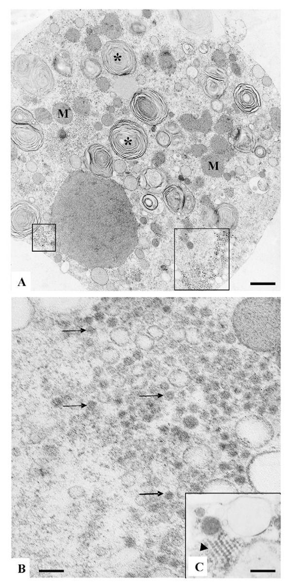
Zika virus (ZIKV) has infected thousands of Brazilian people and spread to other American countries since 2015. The introduction of ZIKV brought a strong impact to public health in Brazil. It is of utmost importance to identify a susceptible cell line that will enable the isolation and identification of the virus from patient samples, viral mass production, and testing of drug and vaccine candidates. Besides real-time reverse transcriptase polymerase chain reaction diagnosis for detecting the viral genome, virus isolation in cell lines was useful in order to study the structure of the viral particle and its behaviour inside cells. Analysis of ZIKV infected cell lines was achieved using transmission electron microscopy (TEM). Blood was obtained from a Brazilian patient during the first days after presenting with signs of the disease, and ZIKV from the patient's blood was isolated in the C6/36 mosquito cell line. Afterwards, Vero cells were inoculated with the viral suspension, fixed six days after inoculation, embedded in polymers, and ultra-thin cut. Like dengue viruses, this flavivirus showed numerous virus particles present inside cellular vesicles thereby confirming the susceptibility of the Vero cell line to ZIKV replication. TEM is a unique technique available to make the virus visible.
自2015年以来,寨卡病毒(ZIKV)已感染数千名巴西人并传播到其他美洲国家。寨卡病毒的传入给巴西的公共卫生带来了巨大冲击。确定一种易感细胞系至关重要,这将有助于从患者样本中分离和鉴定病毒、进行病毒大规模生产以及测试候选药物和疫苗。除了用于检测病毒基因组的实时逆转录聚合酶链反应诊断外,在细胞系中进行病毒分离对于研究病毒颗粒的结构及其在细胞内的行为也很有用。使用透射电子显微镜(TEM)对寨卡病毒感染的细胞系进行分析。在一名出现疾病症状后的头几天从一名巴西患者身上采集血液,并在C6/36蚊细胞系中分离出该患者血液中的寨卡病毒。随后,用病毒悬液接种Vero细胞,并在接种六天后固定,包埋在聚合物中,然后进行超薄切片。与登革病毒一样,这种黄病毒在细胞囊泡内显示出大量病毒颗粒,从而证实了Vero细胞系对寨卡病毒复制的敏感性。透射电子显微镜是一种能使病毒可见的独特技术。