Terry Louise, Cassels Nicola, Lu Kelly, Acton Jennifer H, Margrain Tom H, North Rachel V, Fergusson James, White Nick, Wood Ashley
School of Optometry and Vision Sciences, Cardiff University, Cardiff, United Kingdom.
Vision Science Bioimaging Labs, Cardiff University, Cardiff, United Kingdom.
PLoS One. 2016 Sep 2;11(9):e0162001. doi: 10.1371/journal.pone.0162001. eCollection 2016.
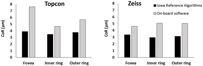
Retinal and intra-retinal layer thicknesses are routinely generated from optical coherence tomography (OCT) images, but on-board software capabilities and image scaling assumptions are not consistent across devices. This study evaluates the device-independent Iowa Reference Algorithms (Iowa Institute for Biomedical Imaging) for automated intra-retinal layer segmentation and image scaling for three OCT systems. Healthy participants (n = 25) underwent macular volume scans using a Cirrus HD-OCT (Zeiss), 3D-OCT 1000 (Topcon), and a non-commercial long-wavelength (1040nm) OCT on two occasions. Mean thickness of 10 intra-retinal layers was measured in three ETDRS subfields (fovea, inner ring and outer ring) using the Iowa Reference Algorithms. Where available, total retinal thicknesses were measured using on-board software. Measured axial eye length (AEL)-dependent scaling was used throughout, with a comparison made to the system-specific fixed-AEL scaling. Inter-session repeatability and agreement between OCT systems and segmentation methods was assessed. Inter-session coefficient of repeatability (CoR) for the foveal subfield total retinal thickness was 3.43μm, 4.76μm, and 5.98μm for the Zeiss, Topcon, and long-wavelength images respectively. For the commercial software, CoR was 4.63μm (Zeiss) and 7.63μm (Topcon). The Iowa Reference Algorithms demonstrated higher repeatability than the on-board software and, in addition, reliably segmented all 10 intra-retinal layers. With fixed-AEL scaling, the algorithm produced significantly different thickness values for the three OCT devices (P<0.05), with these discrepancies generally characterized by an overall offset (bias) and correlations with axial eye length for the foveal subfield and outer ring (P<0.05). This correlation was reduced to an insignificant level in all cases when AEL-dependent scaling was used. Overall, the Iowa Reference Algorithms are viable for clinical and research use in healthy eyes imaged with these devices, however ocular biometry is required for accurate quantification of OCT images.
视网膜及视网膜内各层厚度通常由光学相干断层扫描(OCT)图像生成,但不同设备的机载软件功能及图像缩放假设并不一致。本研究评估了用于三种OCT系统的、与设备无关的爱荷华参考算法(爱荷华生物医学成像研究所),以实现视网膜内各层的自动分割及图像缩放。25名健康受试者分两次使用Cirrus HD-OCT(蔡司)、3D-OCT 1000(拓普康)以及一台非商用长波长(1040nm)OCT进行黄斑体积扫描。使用爱荷华参考算法在三个ETDRS子区域(中央凹、内环和外环)测量10个视网膜内层的平均厚度。如有可用数据,使用机载软件测量总视网膜厚度。全程采用与测量的眼轴长度(AEL)相关的缩放,并与系统特定的固定AEL缩放进行比较。评估了OCT系统与分割方法之间的组间重复性及一致性。中央凹子区域总视网膜厚度的组间重复性系数(CoR),蔡司、拓普康和长波长图像分别为3.43μm、4.76μm和5.98μm。对于商用软件,CoR分别为4.63μm(蔡司)和7.63μm(拓普康)。爱荷华参考算法显示出比机载软件更高的重复性,此外,还能可靠地分割所有10个视网膜内层。采用固定AEL缩放时,该算法对三种OCT设备得出的厚度值有显著差异(P<0.05),这些差异通常表现为整体偏移(偏差)以及与中央凹子区域和外环的眼轴长度相关(P<0.05)。当使用与AEL相关的缩放时,这种相关性在所有情况下均降至不显著水平。总体而言,爱荷华参考算法对于使用这些设备成像的健康眼睛的临床和研究应用是可行的,然而,准确量化OCT图像需要眼部生物测量。