James P D, Atkinson M
Department of Histopathology and Surgery, University Hospital, Queens Medical Centre, Nottingham.
Gut. 1989 Jul;30(7):899-905. doi: 10.1136/gut.30.7.899.
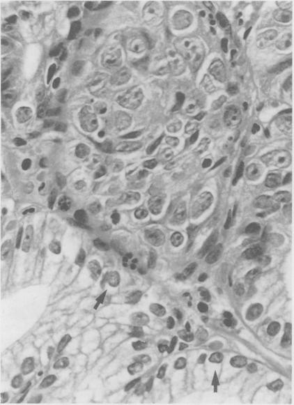
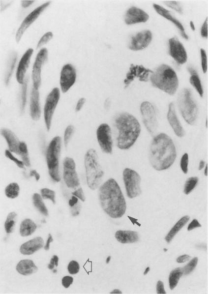
DNA image cytometry was performed on Feulgen stained sections from 91 biopsies obtained during prospective endoscopic surveillance of 55 patients with Barrett's oesophagus. Aneuploid cells were detected in specimens from six of these patients. Four subsequently developed dysplasia and adenocarcinoma but, in the other two, biopsies had been reported as showing specialised epithelium only, with no apparent dysplasia and no evidence of malignancy on clinical follow up to date. In two of the four patients who subsequently developed carcinoma, aneuploid cells were only found in biopsies showing overt dysplasia or carcinoma but in the two other patients aneuploid cells were present in biopsies taken early in the clinical course before any dysplasia had been identified on the original reports. The presence of aneuploid cells on cytometry of these 'benign' biopsies allowed us, on histological review, to identify areas of atypia which were interpreted as mild dysplasia. In this series aneuploidy was always associated with some morphological abnormality varying from mild dysplasia to frank carcinoma. Aneuploid cells were not shown in material from one patient who had an oesophagectomy for dysplasia or in biopsy material from four patients showing 'indefinite dysplasia'. DNA cytometry combines an objective assessment of epithelial atypia with the advantage of detecting rare cellular aneuploidy and the ability to correlate these events with morphology. It should assist in the more accurate diagnosis of dysplasia and prove useful in identifying those patients with Barrett's oesophagus who are at greater risk of subsequently developing malignancy.
对55例巴雷特食管患者进行前瞻性内镜监测期间获取的91份活检组织的福尔根染色切片进行了DNA图像细胞计数分析。在其中6例患者的标本中检测到非整倍体细胞。4例随后发展为发育异常和腺癌,但另外2例的活检报告仅显示为特殊上皮,无明显发育异常,且截至目前的临床随访无恶性肿瘤证据。在随后发展为癌的4例患者中,2例仅在显示明显发育异常或癌的活检组织中发现非整倍体细胞,而另外2例患者在临床病程早期采集的活检组织中就存在非整倍体细胞,当时原始报告中尚未发现任何发育异常。对这些“良性”活检组织进行细胞计数分析时发现存在非整倍体细胞,这使我们在组织学复查时能够识别出被解释为轻度发育异常的异型区域。在该系列研究中,非整倍体总是与一些形态学异常相关,从轻度发育异常到明显的癌不等。1例因发育异常接受食管切除术的患者的材料以及4例显示“不确定发育异常”的患者的活检材料中均未显示非整倍体细胞。DNA细胞计数分析将上皮异型性的客观评估与检测罕见细胞非整倍体的优势以及将这些情况与形态学相关联的能力结合起来。它应有助于更准确地诊断发育异常,并证明对识别那些随后发生恶性肿瘤风险更高的巴雷特食管患者有用。