• 文献检索
  • 文档翻译
  • 深度研究
  • 学术资讯
  • Suppr Zotero 插件Zotero 插件
  • 邀请有礼
  • 套餐&价格
  • 历史记录
应用&插件
Suppr Zotero 插件Zotero 插件浏览器插件Mac 客户端Windows 客户端微信小程序
定价
高级版会员购买积分包购买API积分包
服务
文献检索文档翻译深度研究API 文档MCP 服务
关于我们
关于 Suppr公司介绍联系我们用户协议隐私条款
关注我们

Suppr 超能文献

核心技术专利:CN118964589B侵权必究
粤ICP备2023148730 号-1Suppr @ 2026

文献检索

告别复杂PubMed语法,用中文像聊天一样搜索,搜遍4000万医学文献。AI智能推荐,让科研检索更轻松。

立即免费搜索

文件翻译

保留排版,准确专业,支持PDF/Word/PPT等文件格式,支持 12+语言互译。

免费翻译文档

深度研究

AI帮你快速写综述,25分钟生成高质量综述,智能提取关键信息,辅助科研写作。

立即免费体验

动态对比增强 CT 在胰腺癌患者中的应用。

Dynamic Contrast-Enhanced CT in Patients with Pancreatic Cancer.

机构信息

Department of Diagnostic Radiology, Rigshospitalet, Copenhagen University Hospital, DK-2100 Copenhagen, Denmark.

Department of Technology, Faculty of Health and Technology, Metropolitan University College, DK-2100 Copenhagen, Denmark.

出版信息

Diagnostics (Basel). 2016 Sep 6;6(3):34. doi: 10.3390/diagnostics6030034.

DOI:10.3390/diagnostics6030034
PMID:27608045
原文链接:https://pmc.ncbi.nlm.nih.gov/articles/PMC5039568/
Abstract

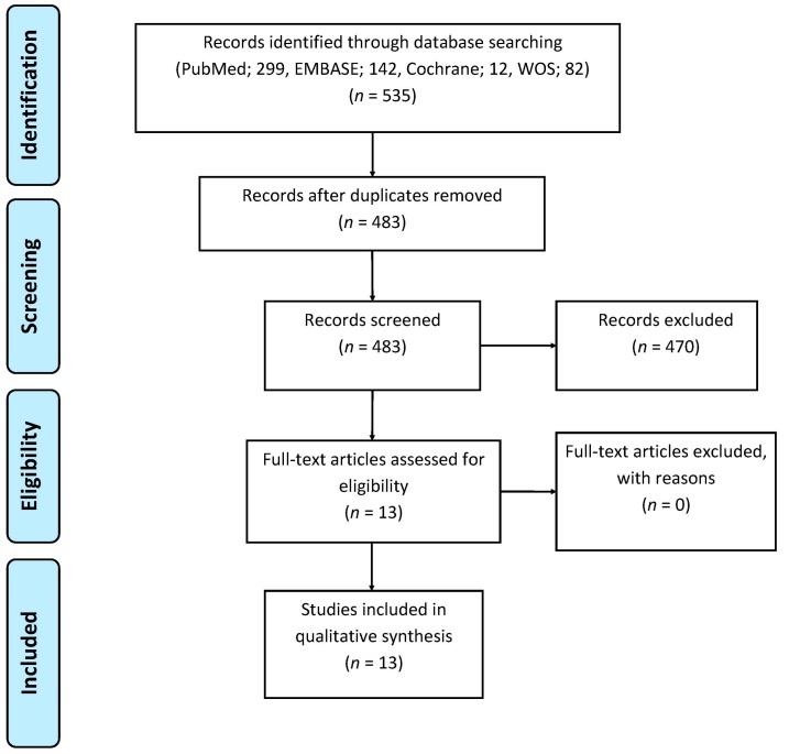
The aim of this systematic review is to provide an overview of the use of Dynamic Contrast-enhanced Computed Tomography (DCE-CT) in patients with pancreatic cancer. This study was composed according to the PRISMA guidelines 2009. The literature search was conducted in PubMed, Cochrane Library, EMBASE, and Web of Science databases to identify all relevant publications. The QUADAS-2 tool was implemented to assess the risk of bias and applicability concerns of each included study. The initial literature search yielded 483 publications. Thirteen articles were included. Articles were categorized into three groups: nine articles concerning primary diagnosis or staging, one article about tumor response to treatment, and three articles regarding scan techniques. In exocrine pancreatic tumors, measurements of blood flow in eight studies and blood volume in seven studies were significantly lower in tumor tissue, compared with measurements in pancreatic tissue outside of tumor, or normal pancreatic tissue in control groups of healthy volunteers. The studies were heterogeneous in the number of patients enrolled and scan protocols. Perfusion parameters measured and analyzed by DCE-CT might be useful in the investigation of characteristic vascular patterns of exocrine pancreatic tumors. Further clinical studies are desired for investigating the potential of DCE-CT in pancreatic tumors.

摘要

本系统评价的目的是提供一个使用动态对比增强计算机断层扫描(DCE-CT)在胰腺癌患者的概述。本研究根据 PRISMA 指南 2009 进行。文献检索在 PubMed、Cochrane 图书馆、EMBASE 和 Web of Science 数据库中进行,以确定所有相关的出版物。采用 QUADAS-2 工具评估每个纳入研究的偏倚风险和适用性问题。最初的文献检索产生了 483 篇出版物。有 13 篇文章被纳入。文章分为三组:9 篇关于原发性诊断或分期,1 篇关于肿瘤对治疗的反应,以及 3 篇关于扫描技术。在外分泌胰腺肿瘤中,与肿瘤外胰腺组织或健康志愿者对照组的正常胰腺组织相比,八项研究中肿瘤组织的血流测量值和七项研究中血容量测量值均显著降低。研究在入组患者数量和扫描方案上存在异质性。DCE-CT 测量和分析的灌注参数可能有助于研究外分泌胰腺肿瘤的特征性血管模式。需要进一步的临床研究来研究 DCE-CT 在胰腺肿瘤中的潜力。

https://cdn.ncbi.nlm.nih.gov/pmc/blobs/f6ba/5039568/4cdddfbf9466/diagnostics-06-00034-g001.jpg

相似文献

1
Dynamic Contrast-Enhanced CT in Patients with Pancreatic Cancer.动态对比增强 CT 在胰腺癌患者中的应用。
Diagnostics (Basel). 2016 Sep 6;6(3):34. doi: 10.3390/diagnostics6030034.
2
Assessing Tumor Response to Treatment in Patients with Lung Cancer Using Dynamic Contrast-Enhanced CT.使用动态对比增强 CT 评估肺癌患者的肿瘤治疗反应。
Diagnostics (Basel). 2016 Jul 21;6(3):28. doi: 10.3390/diagnostics6030028.
3
Dynamic contrast-enhanced micro-computed tomography correlates with 3-dimensional fluorescence ultramicroscopy in antiangiogenic therapy of breast cancer xenografts.动态对比增强微计算机断层扫描与三维荧光超显微镜在乳腺癌异种移植抗血管生成治疗中的相关性。
Invest Radiol. 2014 Jul;49(7):445-56. doi: 10.1097/RLI.0000000000000038.
4
The diagnostic performance of CT versus FDG PET-CT for the detection of recurrent pancreatic cancer: a systematic review and meta-analysis.CT 与 FDG PET-CT 诊断复发胰腺癌的效能比较:系统评价和荟萃分析。
Eur J Radiol. 2018 Sep;106:128-136. doi: 10.1016/j.ejrad.2018.07.010. Epub 2018 Jul 12.
5
Voxelwise comparison of perfusion parameters estimated using dynamic contrast enhanced (DCE) computed tomography and DCE-magnetic resonance imaging in locally advanced cervical cancer.基于动态对比增强 CT 和 DCE 磁共振成像的局部晚期宫颈癌灌注参数的体素比较。
Acta Oncol. 2013 Oct;52(7):1360-8. doi: 10.3109/0284186X.2013.813637. Epub 2013 Sep 5.
6
Development of a dynamic flow imaging phantom for dynamic contrast-enhanced CT.动态对比增强 CT 用动态血流成像模体的研制。
Med Phys. 2011 Aug;38(8):4866-80. doi: 10.1118/1.3615058.
7
Pancreatic CT perfusion: quantitative meta-analysis of disease discrimination, protocol development, and effect of CT parameters.胰腺CT灌注:疾病鉴别、方案制定及CT参数影响的定量荟萃分析
Insights Imaging. 2023 Jul 21;14(1):132. doi: 10.1186/s13244-023-01471-0.
8
Can visual assessment of blood flow patterns by dynamic contrast-enhanced computed tomography distinguish between malignant and benign lung tumors?通过动态对比增强计算机断层扫描对血流模式进行视觉评估能否区分肺恶性肿瘤和良性肿瘤?
Acta Radiol Open. 2017 May 30;6(5):2058460117710053. doi: 10.1177/2058460117710053. eCollection 2017 May.
9
Pancreatic endocrine tumors: tumor blood flow assessed with perfusion CT reflects angiogenesis and correlates with prognostic factors.胰腺内分泌肿瘤:用灌注CT评估的肿瘤血流反映血管生成并与预后因素相关。
Radiology. 2009 Feb;250(2):407-16. doi: 10.1148/radiol.2501080291. Epub 2008 Dec 18.
10
Diagnostic accuracy of flat-panel computed tomography in assessing cerebral perfusion in comparison with perfusion computed tomography and perfusion magnetic resonance: a systematic review.平板计算机断层扫描在评估脑灌注中的诊断准确性与灌注计算机断层扫描和灌注磁共振成像的比较:系统评价。
Neuroradiology. 2019 Dec;61(12):1457-1468. doi: 10.1007/s00234-019-02285-y. Epub 2019 Sep 16.

引用本文的文献

1
Multitasking dynamic contrast enhanced magnetic resonance imaging can accurately differentiate chronic pancreatitis from pancreatic ductal adenocarcinoma.多任务动态对比增强磁共振成像能够准确区分慢性胰腺炎和胰腺导管腺癌。
Front Oncol. 2023 Jan 6;12:1007134. doi: 10.3389/fonc.2022.1007134. eCollection 2022.
2
Combination of DCE-MRI and DWI in Predicting the Treatment Effect of Concurrent Chemoradiotherapy in Esophageal Carcinoma.动态对比增强磁共振成像(DCE-MRI)与扩散加权成像(DWI)联合应用预测食管癌同步放化疗的疗效
Biomed Res Int. 2020 Jun 16;2020:2576563. doi: 10.1155/2020/2576563. eCollection 2020.
3
Fibrotic Response to Neoadjuvant Therapy Predicts Survival in Pancreatic Cancer and Is Measurable with Collagen-Targeted Molecular MRI.

本文引用的文献

1
Computed Tomography (CT) Perfusion in Abdominal Cancer: Technical Aspects.腹部癌症的计算机断层扫描 (CT) 灌注:技术方面。
Diagnostics (Basel). 2013 Apr 3;3(2):261-70. doi: 10.3390/diagnostics3020261.
2
Cancer of the pancreas: ESMO Clinical Practice Guidelines for diagnosis, treatment and follow-up.胰腺癌:ESMO诊断、治疗及随访临床实践指南
Ann Oncol. 2015 Sep;26 Suppl 5:v56-68. doi: 10.1093/annonc/mdv295.
3
Reproducibility of VPCT parameters in the normal pancreas: comparison of two different kinetic calculation models.
新辅助治疗后的纤维化反应可预测胰腺癌的生存情况,并可通过胶原靶向分子 MRI 进行测量。
Clin Cancer Res. 2020 Sep 15;26(18):5007-5018. doi: 10.1158/1078-0432.CCR-18-1359. Epub 2020 Jul 1.
正常胰腺中VPCT参数的可重复性:两种不同动力学计算模型的比较
Acad Radiol. 2015 Sep;22(9):1099-105. doi: 10.1016/j.acra.2015.04.005. Epub 2015 Jun 23.
4
The primary study of low-dose pancreas perfusion by 640- slice helical CT: a whole-organ perfusion.640层螺旋CT低剂量胰腺灌注的初步研究:全器官灌注
Springerplus. 2015 Apr 21;4:192. doi: 10.1186/s40064-015-0950-6. eCollection 2015.
5
Perfusion in the tissue surrounding pancreatic cancer and the patient's prognosis.胰腺癌周围组织的灌注与患者预后。
Biomed Res Int. 2014;2014:648021. doi: 10.1155/2014/648021. Epub 2014 Sep 11.
6
Imaging of pancreatic neoplasms.胰腺肿瘤的影像学检查
Surg Oncol Clin N Am. 2014 Oct;23(4):751-88. doi: 10.1016/j.soc.2014.07.002.
7
Imaging diagnosis of pancreatic cancer: a state-of-the-art review.胰腺癌的影像学诊断:最新综述
World J Gastroenterol. 2014 Jun 28;20(24):7864-77. doi: 10.3748/wjg.v20.i24.7864.
8
Functional imaging in lung cancer.肺癌中的功能成像
Clin Physiol Funct Imaging. 2014 Sep;34(5):340-55. doi: 10.1111/cpf.12104. Epub 2013 Dec 1.
9
Low-dose whole organ CT perfusion of the pancreas: preliminary study.胰腺低剂量全器官CT灌注:初步研究。
Abdom Imaging. 2014 Feb;39(1):40-7. doi: 10.1007/s00261-013-0045-1.
10
Diagnostic value of CT perfusion imaging for parotid neoplasms.CT 灌注成像对腮腺肿瘤的诊断价值。
Dentomaxillofac Radiol. 2014;43(1):20130237. doi: 10.1259/dmfr.20130237. Epub 2013 Nov 1.