Donahue Manus J, Juttukonda Meher R, Watchmaker Jennifer M
Radiology and Radiological Sciences, Vanderbilt University School of Medicine, Nashville, TN, USA; Neurology, Vanderbilt University School of Medicine, Nashville, TN, USA; Psychiatry, Vanderbilt University School of Medicine, Nashville, TN, USA.
Radiology and Radiological Sciences, Vanderbilt University School of Medicine, Nashville, TN, USA.
Neuroimage. 2017 Jul 1;154:43-58. doi: 10.1016/j.neuroimage.2016.09.007. Epub 2016 Sep 11.
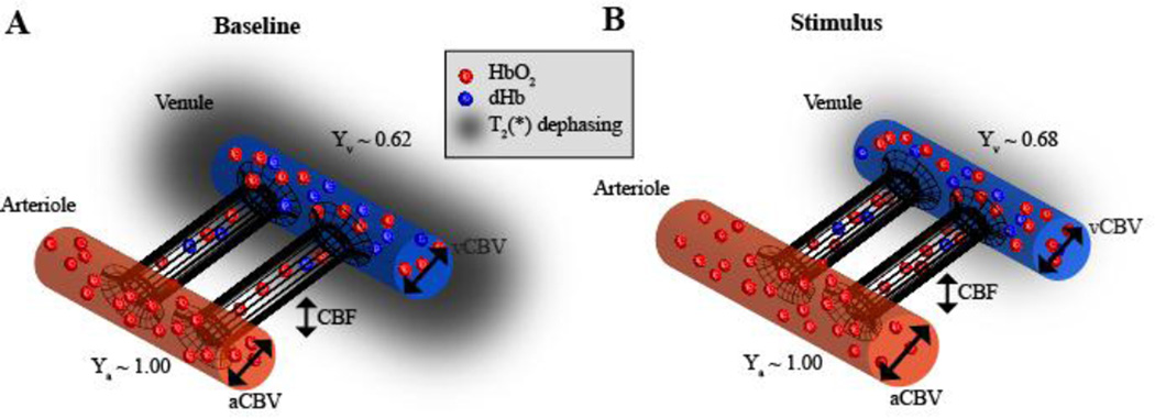
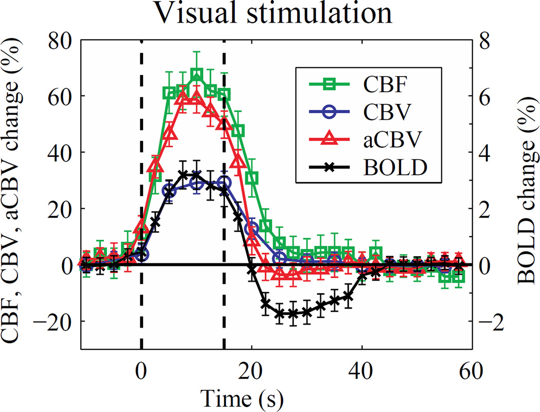
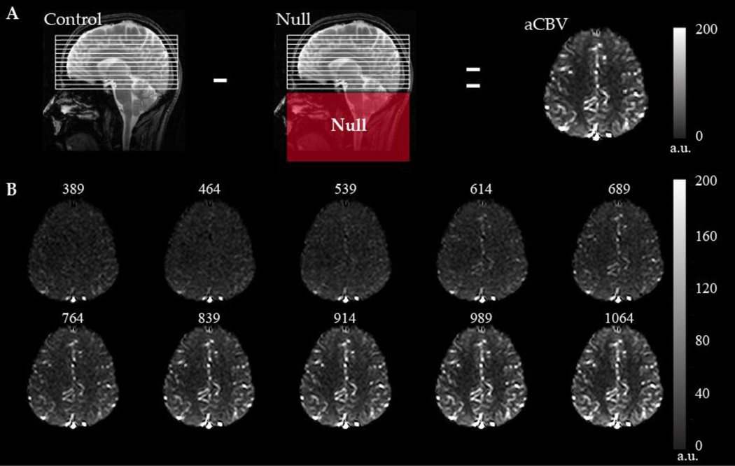
Functional neuroimaging with blood oxygenation level-dependent (BOLD) contrast has emerged as the most popular method for evaluating qualitative changes in brain function in humans. At typical human field strengths (1.5-3.0T), BOLD contrast provides a measure of changes in transverse water relaxation rates in and around capillary and venous blood, and as such provides only a surrogate marker of brain function that depends on dynamic changes in hemodynamics (e.g., cerebral blood flow and volume) and metabolism (e.g., oxygen extraction fraction and the cerebral metabolic rate of oxygen consumption). Alternative functional neuroimaging methods that are specifically sensitive to these constituents of the BOLD signal are being developed and applied in a growing number of clinical and neuroscience applications of quantitative cerebral physiology. These methods require additional considerations for interpreting and quantifying their contrast responsibly. Here, an overview of two popular methods, arterial spin labeling and vascular space occupancy, is presented specifically in the context of functional neuroimaging. Appropriate post-processing and experimental acquisition strategies are summarized with the motivation of reducing sensitivity to noise and unintended signal sources and improving quantitative accuracy of cerebral hemodynamics.
利用血氧水平依赖(BOLD)对比的功能神经成像已成为评估人类脑功能定性变化最常用的方法。在典型的人体场强(1.5 - 3.0T)下,BOLD对比提供了一种测量毛细血管和静脉血内及周围横向水弛豫率变化的方法,因此它仅提供了一种依赖于血流动力学(如脑血流量和血容量)和代谢(如氧摄取分数和脑氧代谢率)动态变化的脑功能替代标志物。正在开发并将对BOLD信号这些成分具有特异性敏感性的替代功能神经成像方法应用于越来越多的定量脑生理学临床和神经科学应用中。这些方法在负责任地解释和量化其对比方面需要额外考虑。在此,特别在功能神经成像的背景下,对两种常用方法——动脉自旋标记和血管空间占据进行概述。总结了适当的后处理和实验采集策略,目的是降低对噪声和非预期信号源的敏感性,并提高脑血流动力学的定量准确性。