Minguillon Jesus, Lopez-Gordo Miguel A, Pelayo Francisco
Department of Computer Architecture and Technology, University of GranadaGranada, Spain; Research Centre for Information and Communications Technologies, University of GranadaGranada, Spain.
Department of Signal Theory, Telematics and Communications, University of GranadaGranada, Spain; Nicolo AssociationGranada, Spain.
Front Comput Neurosci. 2016 Sep 22;10:101. doi: 10.3389/fncom.2016.00101. eCollection 2016.
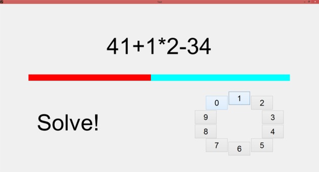
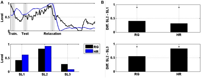
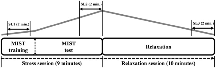
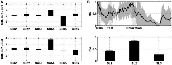
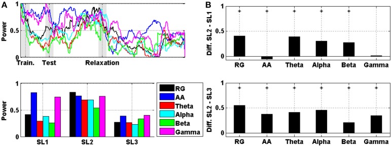
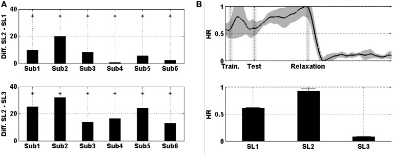
Stress assessment has been under study in the last years. Both biochemical and physiological markers have been used to measure stress level. In neuroscience, several studies have related modification of stress level to brain activity changes in limbic system and frontal regions, by using non-invasive techniques such as functional magnetic resonance imaging (fMRI) and electroencephalography (EEG). In particular, previous studies suggested that the exhibition or inhibition of certain brain rhythms in frontal cortical areas indicates stress. However, there is no established marker to measure stress level by EEG. In this work, we aimed to prove the usefulness of the prefrontal relative gamma power (RG) for stress assessment. We conducted a study based on stress and relaxation periods. Six healthy subjects performed the Montreal Imaging Stress Task (MIST) followed by a stay within a relaxation room while EEG and electrocardiographic signals were recorded. Our results showed that the prefrontal RG correlated with the expected stress level and with the heart rate (HR; 0.8). In addition, the difference in prefrontal RG between time periods of different stress level was statistically significant ( < 0.01). Moreover, the RG was more discriminative between stress levels than alpha asymmetry, theta, alpha, beta, and gamma power in prefrontal cortex. We propose the prefrontal RG as a marker for stress assessment. Compared with other established markers such as the HR or the cortisol, it has higher temporal resolution. Additionally, it needs few electrodes located at non-hairy head positions, thus facilitating the use of non-invasive dry wearable real-time devices for ubiquitous assessment of stress.
近年来,压力评估一直处于研究之中。生化和生理标志物均已被用于测量压力水平。在神经科学领域,通过使用功能磁共振成像(fMRI)和脑电图(EEG)等非侵入性技术,多项研究已将压力水平的变化与边缘系统和额叶区域的大脑活动变化联系起来。特别是,先前的研究表明额叶皮质区域某些脑电波节律的出现或抑制表明存在压力。然而,尚无通过脑电图测量压力水平的确立标志物。在这项工作中,我们旨在证明前额叶相对伽马功率(RG)对压力评估的有用性。我们进行了一项基于压力期和放松期的研究。六名健康受试者执行了蒙特利尔成像压力任务(MIST),随后待在一个放松室中,同时记录脑电图和心电图信号。我们的结果表明,前额叶RG与预期的压力水平以及心率(HR;0.8)相关。此外,不同压力水平时间段之间前额叶RG的差异具有统计学意义(<0.01)。而且,在区分压力水平方面,RG比前额叶皮质中的阿尔法不对称性、θ波、阿尔法波、贝塔波和伽马功率更具辨别力。我们提出将前额叶RG作为压力评估的标志物。与心率或皮质醇等其他已确立的标志物相比,它具有更高的时间分辨率。此外,它只需要很少的电极位于头部无毛发的位置,从而便于使用非侵入性干式可穿戴实时设备进行压力的普遍评估。