Drebert Zuzanna, MacAskill Mark, Doughty-Shenton Dahlia, De Bosscher Karolien, Bracke Marc, Hadoke Patrick W F, Beck Ilse M
Laboratory of Experimental Cancer Research, Department of Radiation Oncology & Experimental Cancer Research, Ghent University, Ghent, Belgium; Cancer Research Institute Ghent (CRIG), Ghent, Belgium.
University/BHF Centre for Cardiovascular Science, The Queen's Medical Research Institute, University of Edinburgh, Edinburgh, United Kingdom.
Vascul Pharmacol. 2017 Feb;89:19-30. doi: 10.1016/j.vph.2016.10.004. Epub 2016 Oct 4.
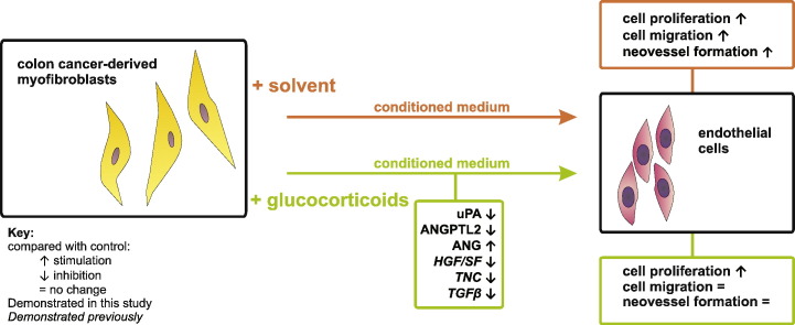
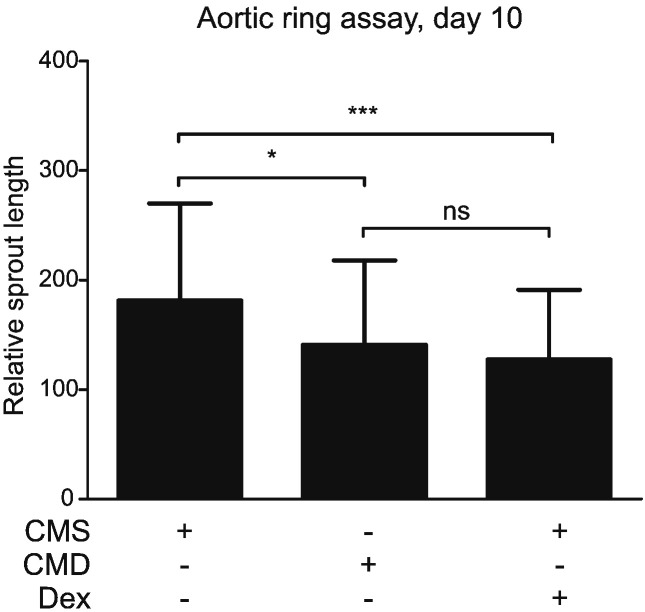
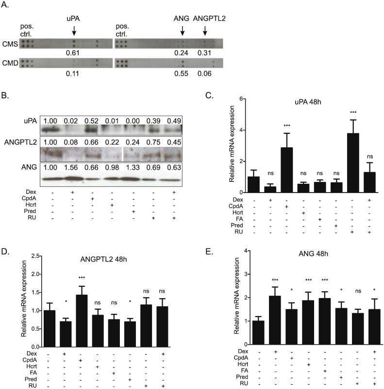
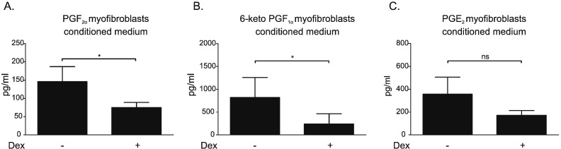
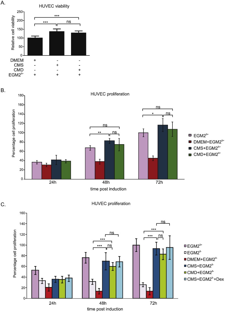
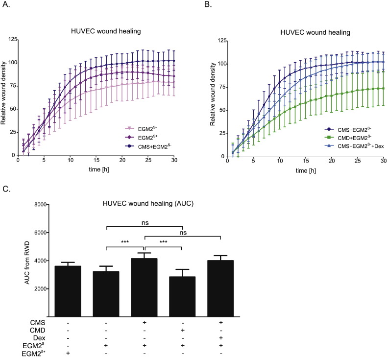
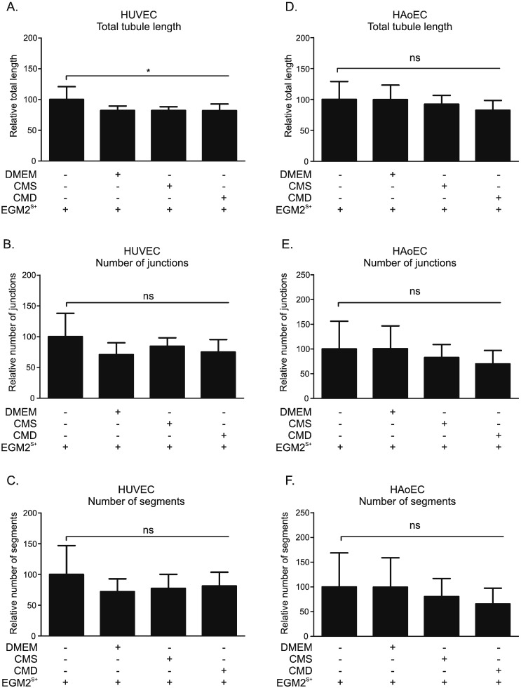
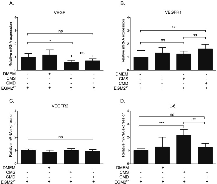
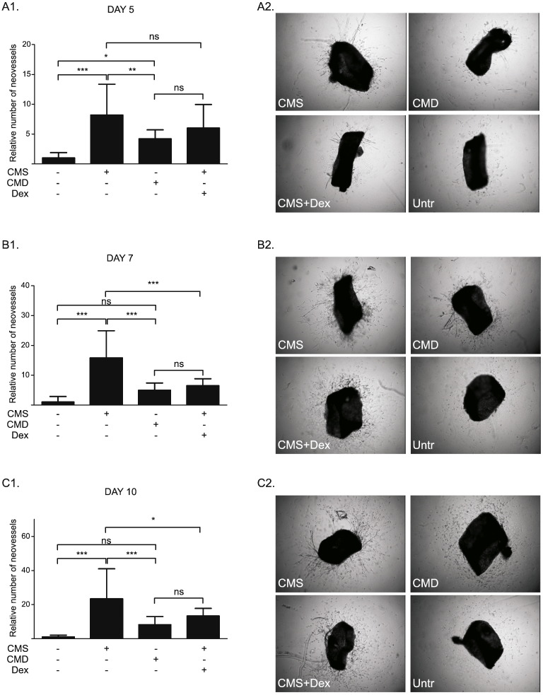
Angiogenesis is important in cancer progression and can be influenced by tumor-associated myofibroblasts. We addressed the hypothesis that glucocorticoids indirectly affect angiogenesis by altering the release of pro-angiogenic factors from colon cancer-derived myofibroblasts. Our study shows that glucocorticoids reduced prostanoids, urokinase-type plasminogen activator (uPA) and angiopoietin-like protein-2 (ANGPTL2) levels, but increased angiogenin (ANG) in supernatant from human CT5.3hTERT colon cancer-derived myofibroblasts. Conditioned medium from solvent- (CMS) and dexamethasone (Dex)-treated (CMD) myofibroblasts increased human umbilical vein endothelial cell (HUVEC) proliferation, but did not affect expression of pro-angiogenic factors or tube-like structure formation (by HUVECs or human aortic ECs). In a HUVEC scratch assay CMS-induced acceleration of wound healing was blunted by CMD treatment. Moreover, CMS-induced neovessel growth in mouse aortic rings ex vivo was also blunted using CMD. The latter effect could be ascribed to both Dex-driven reduction of secreted factors and potential residual Dex present in CMD (indicated using a dexamethasone-spiked CMS control). A similar control in the scratch assay, however, revealed that altered levels of factors in the CMD, and not potential residual Dex, were responsible for decreased wound closure. In conclusion, our results suggest that glucocorticoids indirectly alter endothelial cell function during tumor development in vivo.
血管生成在癌症进展中至关重要,且会受到肿瘤相关肌成纤维细胞的影响。我们探讨了糖皮质激素通过改变结肠癌来源的肌成纤维细胞促血管生成因子的释放来间接影响血管生成的假说。我们的研究表明,糖皮质激素降低了人CT5.3hTERT结肠癌来源的肌成纤维细胞上清液中前列腺素、尿激酶型纤溶酶原激活剂(uPA)和血管生成素样蛋白2(ANGPTL2)的水平,但增加了血管生成素(ANG)的水平。来自溶剂处理(CMS)和地塞米松(Dex)处理(CMD)的肌成纤维细胞的条件培养基可增加人脐静脉内皮细胞(HUVEC)的增殖,但不影响促血管生成因子的表达或管状结构形成(由HUVECs或人主动脉内皮细胞形成)。在HUVEC划痕试验中,CMD处理减弱了CMS诱导的伤口愈合加速。此外,在体外,CMD也减弱了CMS诱导的小鼠主动脉环新血管生长。后一种效应可归因于Dex驱动的分泌因子减少以及CMD中存在的潜在残留Dex(使用加用地塞米松的CMS对照表明)。然而,在划痕试验中的类似对照显示,CMD中因子水平的改变而非潜在的残留Dex导致伤口闭合减少。总之,我们的结果表明,糖皮质激素在体内肿瘤发展过程中间接改变内皮细胞功能。