Kamau Anthony Ndirangu, Park Jung-Eun, Park Eui-Soon, Yu Jung-Eun, Rho Jaerang, Shin Hyun-Jin
Laboratory of Infectious Diseases, College of Veterinary Medicine, Chungnam National University, 220 Gungdong, Yuseong, Daejeon, 305-764, Republic of Korea.
Department of Microbiology & Molecular Biology College of Bioscience & Biotechnology, 220 Gungdong, Yuseong, Daejeon, 305-764, Republic of Korea.
Virus Res. 2017 Jan 2;227:150-157. doi: 10.1016/j.virusres.2016.10.004. Epub 2016 Oct 11.
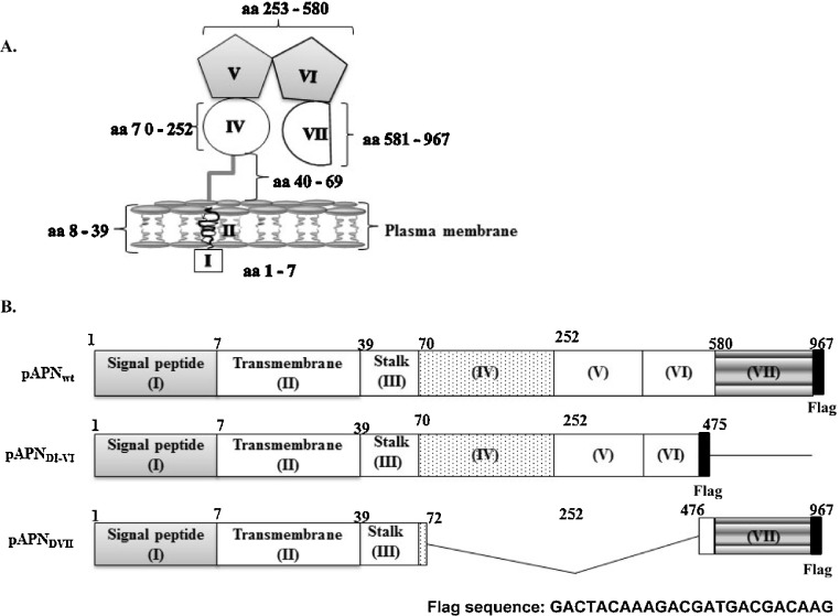
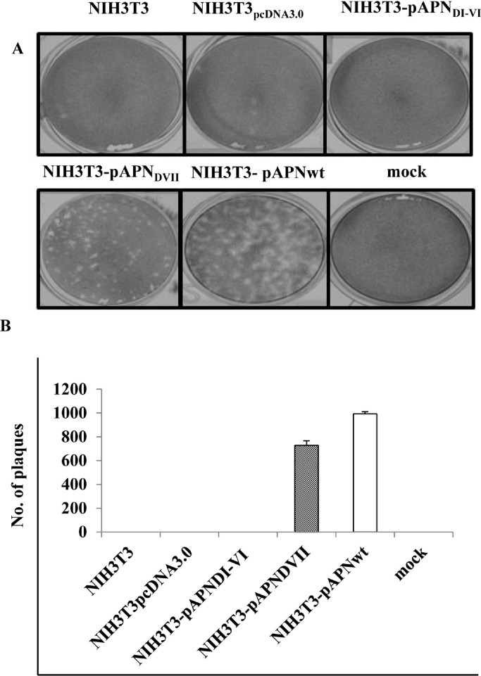
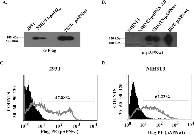
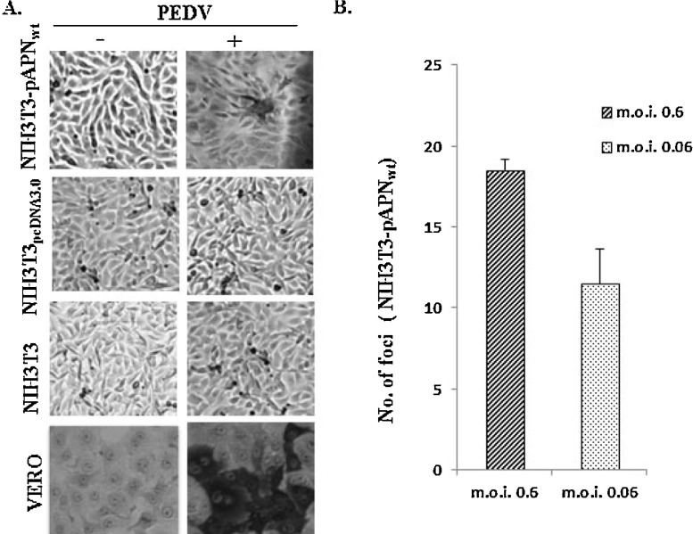
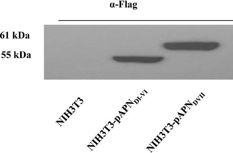
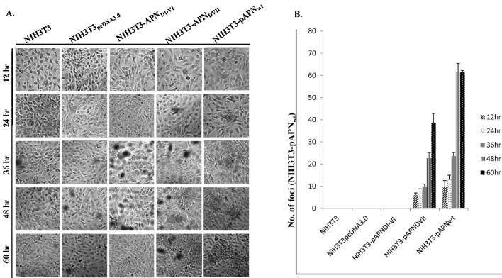
Porcine epidemic diarrhea virus (PEDV) infects swine intestinal cells causing enteric disease. Research has shown that the entry into these cells is through porcine aminopeptidase N (pAPN) receptor. To gain insights into mechanisms of PEDV-pAPN interactions, the present study aimed at identifying the domain that is critical for PEDV binding. To this end, NIH3T3 cell lines constitutively expressing pAPN or pAPN mutants were generated. The mutants were; domain VII deletion mutant and domains IV-VI deletion mutant. In the latter, domain VII was linked to the transmembrane segment through domain III. Results showed PEDV infection was restricted to pAPN and pAPN domain VII expressing NIH3T3 cells. Further, reducing PEDV titre 10 fold resulted in 37.8% decrease in foci indicating positive correlation. A time course test at 12, 24, 36, 48 and 60h showed that foci increased 6 fold in the overall time range. Also, PEDV harvested from pAPN or domain VII expressing NIH3T3 cells was induced indirect plaques in Vero cells confirming successful entry and replication. Collectively, our results demonstrate that PEDV recognizes pAPN and that the main interactive point is lodged within domain VII of the pAPN. These findings are important for therapeutic development as well as creating a platform for future studies on PEDV.
猪流行性腹泻病毒(PEDV)感染猪肠道细胞,引发肠道疾病。研究表明,该病毒进入这些细胞是通过猪氨肽酶N(pAPN)受体。为深入了解PEDV与pAPN的相互作用机制,本研究旨在确定对PEDV结合至关重要的结构域。为此,构建了组成性表达pAPN或pAPN突变体的NIH3T3细胞系。这些突变体包括:结构域VII缺失突变体和结构域IV-VI缺失突变体。在后者中,结构域VII通过结构域III与跨膜区段相连。结果显示,PEDV感染仅限于表达pAPN和pAPN结构域VII的NIH3T3细胞。此外,将PEDV滴度降低10倍导致病灶减少37.8%,表明存在正相关。在12、24、36、48和60小时进行的时间进程测试表明,在整个时间范围内病灶增加了6倍。同样,从表达pAPN或结构域VII的NIH3T3细胞中收获的PEDV在Vero细胞中诱导产生间接空斑,证实其成功进入并复制。总体而言,我们的结果表明PEDV识别pAPN,且主要相互作用点位于pAPN的结构域VII内。这些发现对于治疗性开发以及为未来PEDV研究创建一个平台具有重要意义。