• 文献检索
  • 文档翻译
  • 深度研究
  • 学术资讯
  • Suppr Zotero 插件Zotero 插件
  • 邀请有礼
  • 套餐&价格
  • 历史记录
应用&插件
Suppr Zotero 插件Zotero 插件浏览器插件Mac 客户端Windows 客户端微信小程序
定价
高级版会员购买积分包购买API积分包
服务
文献检索文档翻译深度研究API 文档MCP 服务
关于我们
关于 Suppr公司介绍联系我们用户协议隐私条款
关注我们

Suppr 超能文献

核心技术专利:CN118964589B侵权必究
粤ICP备2023148730 号-1Suppr @ 2026

文献检索

告别复杂PubMed语法,用中文像聊天一样搜索,搜遍4000万医学文献。AI智能推荐,让科研检索更轻松。

立即免费搜索

文件翻译

保留排版,准确专业,支持PDF/Word/PPT等文件格式,支持 12+语言互译。

免费翻译文档

深度研究

AI帮你快速写综述,25分钟生成高质量综述,智能提取关键信息,辅助科研写作。

立即免费体验

结缔组织病患者心肌受累情况的评估:一项多参数心血管磁共振研究。

Evaluation of myocardial involvement in patients with connective tissue disorders: a multi-parametric cardiovascular magnetic resonance study.

作者信息

Mayr Agnes, Kitterer Daniel, Latus Joerg, Steubing Hannah, Henes Joerg, Vecchio Francesco, Kaesemann Philipp, Patrascu Alexandru, Greiser Andreas, Groeninger Stefan, Braun Niko, Alscher M Dominik, Sechtem Udo, Mahrholdt Heiko, Greulich Simon

机构信息

Division of Radiology, University Hospital Innsbruck, Innsbruck, Austria.

Division of Nephrology, Department of Internal Medicine, Robert-Bosch-Medical Center Stuttgart, Stuttgart, Germany.

出版信息

J Cardiovasc Magn Reson. 2016 Oct 13;18(1):67. doi: 10.1186/s12968-016-0288-4.

DOI:10.1186/s12968-016-0288-4
PMID:27733210
原文链接:https://pmc.ncbi.nlm.nih.gov/articles/PMC5062828/
Abstract

BACKGROUND

Severe arrhythmias or heart failure may be surrogates of myocardial involvement in patients with connective tissue disorders (CTD). However, most patients present with unspecific symptoms, normal ECG, and preserved left ventricular ejection fraction (LV-EF). Therefore, timely diagnosis by an accurate technique is crucial. Late gadolinium enhancement (LGE) cardiovascular magnetic resonance (CMR) has proven value for the detection of focal processes, but due to the often diffuse character of fibrosis/inflammation in CTD patients, CMR mapping techniques might be of incremental value for the assessment of myocardial involvement. Purpose of this study was to evaluate a multi-parametric CMR protocol as a screening tool for myocardial involvement in CTD patients.

METHODS

Forty CTD patients were prospectively enrolled and underwent CMR, twenty healthy volunteers served as control group.

RESULTS

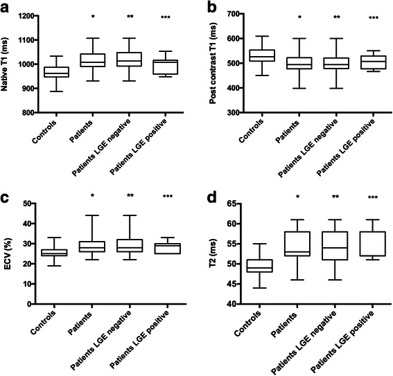
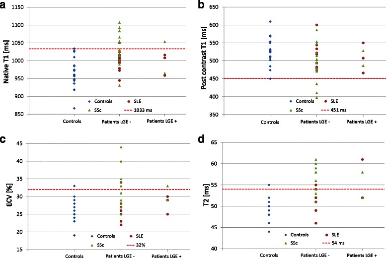
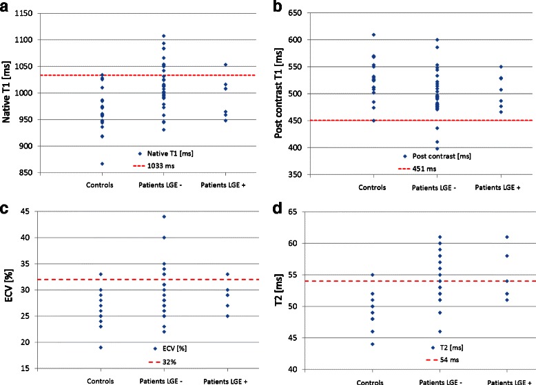
Mean LV-EF was 62 %; LGE prevalence was low (18 %). CTD patients had higher native T1 (1008 vs. 962 ms, p = 0.001), lower post contrast T1 (494 vs. 526 ms, p = 0.008), expanded extracellular volume (ECV) (28 vs. 25 %, p = 0.001), and higher T2 values (53 vs. 49 ms, p < 0.001) compared to controls. Among patients with values higher than the 95 % percentile of healthy controls, native T1 and T2 values seem to be the most promising discriminators.

CONCLUSION

CTD patients showed higher T1, ECV, and T2 values compared to controls, with most significant differences for native T1 and T2, which seem to be independent of the presence of LGE. Our data suggest that CMR mapping techniques are of incremental value in the detection of myocardial involvement in CTD patients.

摘要

背景

严重心律失常或心力衰竭可能是结缔组织病(CTD)患者心肌受累的替代指标。然而,大多数患者表现为非特异性症状、心电图正常且左心室射血分数(LV-EF)保留。因此,采用准确技术进行及时诊断至关重要。延迟钆增强(LGE)心血管磁共振(CMR)已被证明在检测局灶性病变方面具有价值,但由于CTD患者的纤维化/炎症通常具有弥漫性,CMR成像技术可能在评估心肌受累方面具有更大价值。本研究的目的是评估一种多参数CMR方案作为CTD患者心肌受累的筛查工具。

方法

前瞻性纳入40例CTD患者并进行CMR检查,20名健康志愿者作为对照组。

结果

平均LV-EF为62%;LGE患病率较低(18%)。与对照组相比,CTD患者的固有T1值更高(1008对962毫秒,p = 0.001),钆增强后T1值更低(494对526毫秒,p = 0.008),细胞外容积(ECV)扩大(28%对25%,p = 0.001),T2值更高(53对49毫秒,p < 0.001)。在数值高于健康对照者第95百分位数的患者中,固有T1和T2值似乎是最有前景的鉴别指标。

结论

与对照组相比,CTD患者的T1、ECV和T2值更高,固有T1和T2的差异最为显著,且这些差异似乎与LGE的存在无关。我们的数据表明,CMR成像技术在检测CTD患者心肌受累方面具有更大价值。

https://cdn.ncbi.nlm.nih.gov/pmc/blobs/80f6/5062828/6e6526bb3920/12968_2016_288_Fig5_HTML.jpg
https://cdn.ncbi.nlm.nih.gov/pmc/blobs/80f6/5062828/fd10b4305f31/12968_2016_288_Fig1_HTML.jpg
https://cdn.ncbi.nlm.nih.gov/pmc/blobs/80f6/5062828/92cc7651826d/12968_2016_288_Fig2_HTML.jpg
https://cdn.ncbi.nlm.nih.gov/pmc/blobs/80f6/5062828/e2704fbcd8ff/12968_2016_288_Fig3_HTML.jpg
https://cdn.ncbi.nlm.nih.gov/pmc/blobs/80f6/5062828/24b2615df2a4/12968_2016_288_Fig4_HTML.jpg
https://cdn.ncbi.nlm.nih.gov/pmc/blobs/80f6/5062828/6e6526bb3920/12968_2016_288_Fig5_HTML.jpg

相似文献

1
Evaluation of myocardial involvement in patients with connective tissue disorders: a multi-parametric cardiovascular magnetic resonance study.结缔组织病患者心肌受累情况的评估:一项多参数心血管磁共振研究。
J Cardiovasc Magn Reson. 2016 Oct 13;18(1):67. doi: 10.1186/s12968-016-0288-4.
2
T1 and T2 mapping for evaluation of myocardial involvement in patients with ANCA-associated vasculitides.采用T1和T2映射评估抗中性粒细胞胞浆抗体相关性血管炎患者的心肌受累情况。
J Cardiovasc Magn Reson. 2017 Jan 6;19(1):6. doi: 10.1186/s12968-016-0315-5.
3
Diffuse Myocardial Fibrosis and Inflammation in Rheumatoid Arthritis: Insights From CMR T1 Mapping.类风湿关节炎的弥漫性心肌纤维化和炎症:CMR T1 映射的见解。
JACC Cardiovasc Imaging. 2015 May;8(5):526-536. doi: 10.1016/j.jcmg.2014.12.025. Epub 2015 Apr 15.
4
Subclinical myocardial inflammation and diffuse fibrosis are common in systemic sclerosis--a clinical study using myocardial T1-mapping and extracellular volume quantification.系统性硬化症患者存在亚临床心肌炎症和弥漫性纤维化——一项采用心肌 T1 mapping 和细胞外容积定量技术的临床研究。
J Cardiovasc Magn Reson. 2014 Mar 4;16(1):21. doi: 10.1186/1532-429X-16-21.
5
T1 and T2 mapping cardiovascular magnetic resonance imaging techniques reveal unapparent myocardial injury in patients with myocarditis.T1和T2映射心血管磁共振成像技术可揭示心肌炎患者隐匿的心肌损伤。
Clin Res Cardiol. 2017 Jan;106(1):10-17. doi: 10.1007/s00392-016-1018-5. Epub 2016 Jul 7.
6
T-Mapping and extracellular volume estimates in pediatric subjects with Duchenne muscular dystrophy and healthy controls at 3T.3T 下杜氏肌营养不良症患儿与健康对照者的 T 映射和细胞外容积评估。
J Cardiovasc Magn Reson. 2020 Dec 10;22(1):85. doi: 10.1186/s12968-020-00687-z.
7
Advanced myocardial tissue characterisation by a multi-component CMR protocol in patients with rheumatoid arthritis.类风湿关节炎患者应用多组份 CMR 方案进行心肌组织的高级特征分析。
Eur Radiol. 2017 Nov;27(11):4639-4649. doi: 10.1007/s00330-017-4838-4. Epub 2017 May 12.
8
Pattern and prognostic value of cardiac involvement in patients with late-onset pompe disease: a comprehensive cardiovascular magnetic resonance approach.晚发型庞贝病患者心脏受累的模式及预后价值:一种全面的心血管磁共振方法
J Cardiovasc Magn Reson. 2016 Dec 7;18(1):91. doi: 10.1186/s12968-016-0311-9.
9
Cardiac Involvement in Patients Recovered From COVID-2019 Identified Using Magnetic Resonance Imaging.利用磁共振成像技术识别 COVID-19 康复患者的心脏受累情况。
JACC Cardiovasc Imaging. 2020 Nov;13(11):2330-2339. doi: 10.1016/j.jcmg.2020.05.004. Epub 2020 May 12.
10
Native T1-mapping detects the location, extent and patterns of acute myocarditis without the need for gadolinium contrast agents.原生 T1 映射可在无需钆对比剂的情况下检测急性心肌炎的位置、范围和模式。
J Cardiovasc Magn Reson. 2014 May 23;16(1):36. doi: 10.1186/1532-429X-16-36.

引用本文的文献

1
Multiparametric Mapping via Cardiovascular Magnetic Resonance in the Risk Stratification of Ventricular Arrhythmias and Sudden Cardiac Death.多参数心血管磁共振映射在心律失常和心脏性猝死风险分层中的应用。
Medicina (Kaunas). 2024 Apr 24;60(5):691. doi: 10.3390/medicina60050691.
2
Quantitative MRI in cardiometabolic disease: From conventional cardiac and liver tissue mapping techniques to multi-parametric approaches.心脏代谢疾病中的定量磁共振成像:从传统的心脏和肝脏组织成像技术到多参数方法。
Front Cardiovasc Med. 2023 Jan 23;9:991383. doi: 10.3389/fcvm.2022.991383. eCollection 2022.
3
Imaging Methods: Magnetic Resonance Imaging.

本文引用的文献

1
Increased myocardial native T1 and extracellular volume in patients with Duchenne muscular dystrophy.杜兴氏肌营养不良症患者心肌固有T1和细胞外容积增加。
J Cardiovasc Magn Reson. 2016 Jan 21;18:5. doi: 10.1186/s12968-016-0224-7.
2
T1 Mapping in Discrimination of Hypertrophic Phenotypes: Hypertensive Heart Disease and Hypertrophic Cardiomyopathy: Findings From the International T1 Multicenter Cardiovascular Magnetic Resonance Study.T1 Mapping在肥厚型表型鉴别中的应用:高血压性心脏病和肥厚型心肌病:国际T1多中心心血管磁共振研究结果
Circ Cardiovasc Imaging. 2015 Dec;8(12). doi: 10.1161/CIRCIMAGING.115.003285.
3
影像方法:磁共振成像。
Circ Cardiovasc Imaging. 2023 Jan;16(1):e014068. doi: 10.1161/CIRCIMAGING.122.014068. Epub 2023 Jan 17.
4
Utilization of Cardiovascular Magnetic Resonance Imaging for Resumption of Athletic Activities Following COVID-19 Infection: An Expert Consensus Document on Behalf of the American Heart Association Council on Cardiovascular Radiology and Intervention Leadership and Endorsed by the Society for Cardiovascular Magnetic Resonance.COVID-19 感染后恢复运动活动的心血管磁共振成像利用:美国心脏协会心血管放射学和介入领导委员会的专家共识文件,并得到心血管磁共振学会的认可。
Circ Cardiovasc Imaging. 2023 Jan;16(1):e014106. doi: 10.1161/CIRCIMAGING.122.014106. Epub 2022 Dec 21.
5
Utilization of cardiovascular magnetic resonance (CMR) imaging for resumption of athletic activities following COVID-19 infection: an expert consensus document on behalf of the American Heart Association Council on Cardiovascular Radiology and Intervention (CVRI) Leadership and endorsed by the Society for Cardiovascular Magnetic Resonance (SCMR).COVID-19 感染后恢复运动活动的心血管磁共振(CMR)成像的利用:代表美国心脏协会心血管放射学和介入委员会(CVRI)领导层的专家共识文件,并得到心血管磁共振学会(SCMR)的认可。
J Cardiovasc Magn Reson. 2022 Dec 21;24(1):73. doi: 10.1186/s12968-022-00907-8.
6
Using multi-parametric quantitative MRI to screen for cardiac involvement in patients with idiopathic inflammatory myopathy.采用多参数定量 MRI 筛查特发性炎性肌病患者的心脏受累情况。
Sci Rep. 2022 Jun 14;12(1):9819. doi: 10.1038/s41598-022-13858-y.
7
T2 mapping in myocardial disease: a comprehensive review.心肌疾病的 T2 映射:全面综述。
J Cardiovasc Magn Reson. 2022 Jun 6;24(1):33. doi: 10.1186/s12968-022-00866-0.
8
Quantitative assessment of left ventricular myocardial involvement in patients with connective tissue disease: a 3.0T contrast-enhanced cardiovascular magnetic resonance study.结缔组织病患者左心室心肌受累的定量评估:一项3.0T对比增强心血管磁共振研究
Int J Cardiovasc Imaging. 2022 Jul;38(7):1545-1554. doi: 10.1007/s10554-022-02539-6. Epub 2022 Mar 13.
9
A Clinical Approach to Multimodality Imaging in Pulmonary Hypertension.肺动脉高压多模态成像的临床应用方法
Front Cardiovasc Med. 2022 Jan 18;8:794706. doi: 10.3389/fcvm.2021.794706. eCollection 2021.
10
Cardiovascular Magnetic Resonance Reveals Cardiac Pathophysiology in Autoimmune Rheumatic Diseases.心血管磁共振成像揭示自身免疫性风湿疾病中的心脏病理生理学
Mediterr J Rheumatol. 2021 Mar 31;32(1):15-20. doi: 10.31138/mjr.32.1.15. eCollection 2021 Mar.
Performance of t1 and t2 mapping cardiovascular magnetic resonance to detect active myocarditis in patients with recent-onset heart failure.
利用T1和T2 mapping心血管磁共振成像检测近期发生心力衰竭患者的活动性心肌炎
Circ Cardiovasc Imaging. 2015 Jun;8(6). doi: 10.1161/CIRCIMAGING.114.003073.
4
Clinical and histopathological features of patients with systemic sclerosis undergoing endomyocardial biopsy.接受心内膜心肌活检的系统性硬化症患者的临床和组织病理学特征
PLoS One. 2015 May 12;10(5):e0126707. doi: 10.1371/journal.pone.0126707. eCollection 2015.
5
Diffuse Myocardial Fibrosis and Inflammation in Rheumatoid Arthritis: Insights From CMR T1 Mapping.类风湿关节炎的弥漫性心肌纤维化和炎症:CMR T1 映射的见解。
JACC Cardiovasc Imaging. 2015 May;8(5):526-536. doi: 10.1016/j.jcmg.2014.12.025. Epub 2015 Apr 15.
6
Myocardial Inflammation-Are We There Yet?心肌炎症——我们到那一步了吗?
Curr Cardiovasc Imaging Rep. 2015;8(3):6. doi: 10.1007/s12410-015-9320-6.
7
Myocardial T2 mapping by cardiovascular magnetic resonance reveals subclinical myocardial inflammation in patients with systemic lupus erythematosus.通过心血管磁共振进行心肌T2映射揭示了系统性红斑狼疮患者的亚临床心肌炎症。
Int J Cardiovasc Imaging. 2015 Feb;31(2):389-97. doi: 10.1007/s10554-014-0560-3. Epub 2014 Oct 29.
8
Subclinical myocardial inflammation and diffuse fibrosis are common in systemic sclerosis--a clinical study using myocardial T1-mapping and extracellular volume quantification.系统性硬化症患者存在亚临床心肌炎症和弥漫性纤维化——一项采用心肌 T1 mapping 和细胞外容积定量技术的临床研究。
J Cardiovasc Magn Reson. 2014 Mar 4;16(1):21. doi: 10.1186/1532-429X-16-21.
9
Myocardial T1 mapping and extracellular volume quantification: a Society for Cardiovascular Magnetic Resonance (SCMR) and CMR Working Group of the European Society of Cardiology consensus statement.心肌 T1 映射和细胞外容积定量:心血管磁共振学会 (SCMR) 和欧洲心脏病学会 CMR 工作组的共识声明。
J Cardiovasc Magn Reson. 2013 Oct 14;15(1):92. doi: 10.1186/1532-429X-15-92.
10
Cardiac tissue characterization and the diagnostic value of cardiovascular magnetic resonance in systemic connective tissue diseases.心脏组织特征分析及心血管磁共振在系统性结缔组织疾病中的诊断价值。
Arthritis Care Res (Hoboken). 2014 Jan;66(1):104-12. doi: 10.1002/acr.22181.