Pradhan Sulena, Hedberg Jonas, Blomberg Eva, Wold Susanna, Odnevall Wallinder Inger
Division of Surface and Corrosion Science, Department of Chemistry, KTH Royal Institute of Technology, Drottning Kristinas väg 51, 100 44 Stockholm, Sweden.
Division of Surface and Corrosion Science, Department of Chemistry, KTH Royal Institute of Technology, Drottning Kristinas väg 51, 100 44 Stockholm, Sweden ; Chemistry, Materials and Surfaces, SP Technical Research Institute of Sweden, P.O. Box 5607, 114 86 Stockholm, Sweden.
J Nanopart Res. 2016;18(9):285. doi: 10.1007/s11051-016-3597-5. Epub 2016 Sep 22.
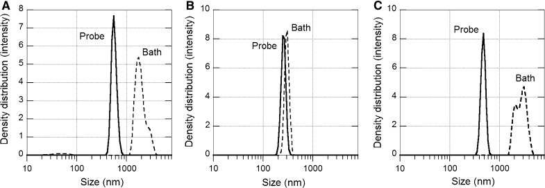
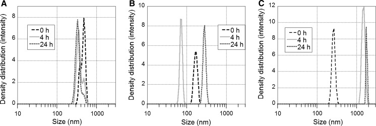
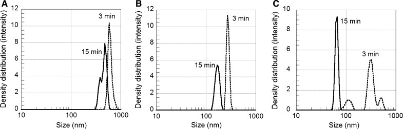
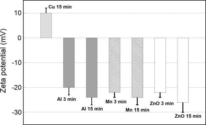
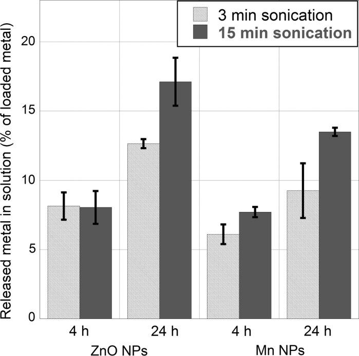
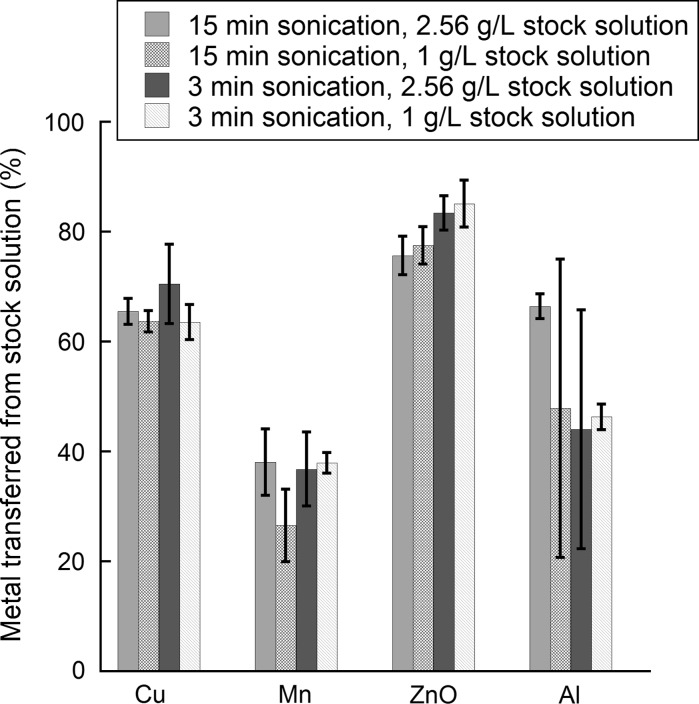
In this study, we elucidate the effect of different sonication techniques to efficiently prepare particle dispersions from selected non-functionalized NPs (Cu, Al, Mn, ZnO), and corresponding consequences on the particle dose, surface charge and release of metals. Probe sonication was shown to be the preferred method for dispersing non-inert, non-functionalized metal NPs (Cu, Mn, Al). However, rapid sedimentation during sonication resulted in differences between the real and the administered doses in the order of 30-80 % when sonicating in 1 and 2.56 g/L NP stock solutions. After sonication, extensive agglomeration of the metal NPs resulted in rapid sedimentation of all particles. DLVO calculations supported these findings, showing the strong van der Waals forces of the metal NPs to result in significant NP agglomeration. Metal release from the metal NPs was slightly increased by increased sonication. The addition of a stabilizing agent (bovine serum albumin) had an accelerating effect on the release of metals in sonicated solutions. For Cu and Mn NPs, the extent of particle dissolution increased from <1.6 to ~5 % after sonication for 15 min. A prolonged sonication time (3-15 min) had negligible effects on the zeta potential of the studied NPs. In all, it is shown that it is of utmost importance to carefully investigate how sonication influences the physico-chemical properties of dispersed metal NPs. This should be considered in nanotoxicology investigations of metal NPs.
在本研究中,我们阐明了不同超声处理技术对从选定的非功能化纳米颗粒(铜、铝、锰、氧化锌)高效制备颗粒分散体的影响,以及对颗粒剂量、表面电荷和金属释放的相应影响。结果表明,探头超声处理是分散非惰性、非功能化金属纳米颗粒(铜、锰、铝)的首选方法。然而,在1和2.56 g/L纳米颗粒储备溶液中进行超声处理时,超声处理过程中的快速沉降导致实际剂量与给药剂量之间的差异在30%-80%左右。超声处理后,金属纳米颗粒的广泛团聚导致所有颗粒迅速沉降。DLVO计算支持了这些发现,表明金属纳米颗粒的强范德华力导致了显著的纳米颗粒团聚。超声处理增加会使金属纳米颗粒的金属释放略有增加。添加稳定剂(牛血清白蛋白)对超声处理溶液中的金属释放有加速作用。对于铜和锰纳米颗粒,超声处理15分钟后,颗粒溶解程度从<1.6%增加到约5%。延长超声处理时间(3-15分钟)对所研究纳米颗粒的zeta电位影响可忽略不计。总之,结果表明,仔细研究超声处理如何影响分散金属纳米颗粒的物理化学性质至关重要。在金属纳米颗粒的纳米毒理学研究中应考虑这一点。