• 文献检索
  • 文档翻译
  • 深度研究
  • 学术资讯
  • Suppr Zotero 插件Zotero 插件
  • 邀请有礼
  • 套餐&价格
  • 历史记录
应用&插件
Suppr Zotero 插件Zotero 插件浏览器插件Mac 客户端Windows 客户端微信小程序
定价
高级版会员购买积分包购买API积分包
服务
文献检索文档翻译深度研究API 文档MCP 服务
关于我们
关于 Suppr公司介绍联系我们用户协议隐私条款
关注我们

Suppr 超能文献

核心技术专利:CN118964589B侵权必究
粤ICP备2023148730 号-1Suppr @ 2026

文献检索

告别复杂PubMed语法,用中文像聊天一样搜索,搜遍4000万医学文献。AI智能推荐,让科研检索更轻松。

立即免费搜索

文件翻译

保留排版,准确专业,支持PDF/Word/PPT等文件格式,支持 12+语言互译。

免费翻译文档

深度研究

AI帮你快速写综述,25分钟生成高质量综述,智能提取关键信息,辅助科研写作。

立即免费体验

不同症状干眼患者睑板腺功能障碍的共聚焦显微镜评估

Confocal Microscopy Evaluation of Meibomian Gland Dysfunction in Dry Eye Patients with Different Symptoms.

作者信息

Zhao Hui, Chen Jing-Yao, Wang Yu-Qian, Lin Zhi-Rong, Wang Shen

机构信息

Eye Institute of Xiamen University, Xiamen, Fujian 361005; Department of Ophthalmology & Visual Science, Medical College of Xiamen University, Xiamen, Fujian 361005; Fujian Provincial Key Laboratory of Ophthalmology and Visual Science, Xiamen, Fujian 361005; Department of Corneal & Ocular Surface Diseases, Xiamen University Affiliated Xiamen Eye Center, Xiamen, Fujian 361005, China.

出版信息

Chin Med J (Engl). 2016 Nov 5;129(21):2617-2622. doi: 10.4103/0366-6999.192782.

DOI:10.4103/0366-6999.192782
PMID:27779170
原文链接:https://pmc.ncbi.nlm.nih.gov/articles/PMC5125342/
Abstract

BACKGROUND

Dry eye patients suffer from all kinds of symptoms. Sometimes, the clinical signs evaluation does not disclose any obvious difference in routine examination; in vivo confocal microscopy (IVCM) is a powerful tool for ocular surface disease. This study aimed to clarify meibomian gland (MG) alterations in dry eye patients with different symptoms and to compare the findings using IVCM.

METHODS

A total of sixty patients were recruited, all subjected to Ocular Surface Disease Index (OSDI) and Salisbury Eye Evaluation Questionnaire (SEEQ), and questionnaires for the assessment of dry eye symptoms before clinical sign examinations were given to the patients. Finally, IVCM was applied to observe MG's structure. Statistical analysis was performed using the t-test, Mann-Whitney U-test and Spearman correlation analysis. The differences were statistically significant when P< 0.05.

RESULTS

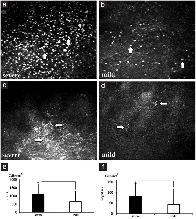
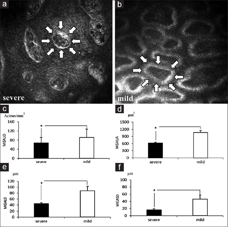
In the severe symptom group, OSDI and SEEQ scores were significantly higher (P< 0.05) compared with the mild symptoms group. All other clinical sign examinations had no statistical difference in the two groups (P> 0.05). However, all the IVCM-observed data showed that patients with severe symptoms had more significant fibrosis in MG (acinar unit area 691.87 ± 182.01 μm2 for the severe, 992.17 ± 170.84 μm2 for the mild; P< 0.05) and severer decrease in the size of MG acinar units than those observed in patients with mild symptoms (MG acinar unit density [MGAUD] 70.08 ± 18.78 glands/mm2, MG acinar unit longest diameter [MGALD] 51.50 ± 15.51 μm, MG acinar unit shortest diameter [MGASD] 20.30 ± 11.85 μm for the severe, MGAUD 89.53 ± 39.88 glands/mm2, MGALD 81.57 ± 21.14 μm, MGASD 42.37 ± 14.55 μm for the mild;P< 0.05). Dry eye symptoms were negatively correlated with MG confocal microscopic parameters and positively correlated with conjunctival inflammatory cells and Langerhans cells (P< 0.05).

CONCLUSIONS

IVCM application provides a strong support to differentiate dry eye patients with different symptoms: meibomian gland dysfunction (MGD) plays a pivotal role in dry eye aggravation, and using IVCM to observe MG fibrosis, changes in size and density of MG as well as status of inflammation cells can help not only correctly diagnose the type and severity of dry eye, but also possibly prognosticate in routine eye examination in the occurrence of MGD.

摘要

背景

干眼症患者会出现各种症状。有时,临床体征评估在常规检查中未发现任何明显差异;共聚焦显微镜检查(IVCM)是诊断眼表疾病的有力工具。本研究旨在明确不同症状的干眼症患者睑板腺(MG)的改变,并使用IVCM比较研究结果。

方法

共招募60例患者,所有患者均接受眼表疾病指数(OSDI)和索尔兹伯里眼评估问卷(SEEQ)评估,并在临床体征检查前向患者发放干眼症症状评估问卷。最后,应用IVCM观察MG的结构。采用t检验、曼-惠特尼U检验和Spearman相关分析进行统计学分析。当P<0.05时,差异具有统计学意义。

结果

与轻度症状组相比,重度症状组的OSDI和SEEQ评分显著更高(P<0.05)。两组间所有其他临床体征检查均无统计学差异(P>0.05)。然而,所有IVCM观察数据均显示,重度症状患者的MG纤维化更明显(重度组腺泡单位面积为691.87±182.01μm²,轻度组为992.17±170.84μm²;P<0.05),且MG腺泡单位大小的减小程度比轻度症状患者更严重(重度组MG腺泡单位密度[MGAUD]为70.08±18.78个腺/mm²,MG腺泡单位最长直径[MGALD]为51.50±15.51μm,MG腺泡单位最短直径[MGASD]为20.30±11.85μm;轻度组MGAUD为89.53±39.88个腺/mm²,MGALD为81.57±21.14μm,MGASD为42.37±14.55μm;P<0.05)。干眼症症状与MG共聚焦显微镜参数呈负相关,与结膜炎症细胞和朗格汉斯细胞呈正相关(P<0.05)。

结论

IVCM的应用为鉴别不同症状的干眼症患者提供了有力支持:睑板腺功能障碍(MGD)在干眼症加重中起关键作用,使用IVCM观察MG纤维化、MG大小和密度变化以及炎症细胞状态不仅有助于正确诊断干眼症的类型和严重程度,还可能在常规眼科检查中对MGD的发生进行预测。

https://cdn.ncbi.nlm.nih.gov/pmc/blobs/f042/5125342/72faa7e12c41/CMJ-129-2617-g003.jpg
https://cdn.ncbi.nlm.nih.gov/pmc/blobs/f042/5125342/fa1f95a18822/CMJ-129-2617-g001.jpg
https://cdn.ncbi.nlm.nih.gov/pmc/blobs/f042/5125342/6ce3fc7878df/CMJ-129-2617-g002.jpg
https://cdn.ncbi.nlm.nih.gov/pmc/blobs/f042/5125342/72faa7e12c41/CMJ-129-2617-g003.jpg
https://cdn.ncbi.nlm.nih.gov/pmc/blobs/f042/5125342/fa1f95a18822/CMJ-129-2617-g001.jpg
https://cdn.ncbi.nlm.nih.gov/pmc/blobs/f042/5125342/6ce3fc7878df/CMJ-129-2617-g002.jpg
https://cdn.ncbi.nlm.nih.gov/pmc/blobs/f042/5125342/72faa7e12c41/CMJ-129-2617-g003.jpg

相似文献

1
Confocal Microscopy Evaluation of Meibomian Gland Dysfunction in Dry Eye Patients with Different Symptoms.不同症状干眼患者睑板腺功能障碍的共聚焦显微镜评估
Chin Med J (Engl). 2016 Nov 5;129(21):2617-2622. doi: 10.4103/0366-6999.192782.
2
[In vivo confocal microscopy evaluation of meibomian glands in meibomian gland dysfunction patients].睑板腺功能障碍患者睑板腺的体内共聚焦显微镜评估
Zhonghua Yan Ke Za Zhi. 2016 Sep 11;52(9):649-56. doi: 10.3760/cma.j.issn.0412-4081.2016.09.004.
3
The efficacy, sensitivity, and specificity of in vivo laser confocal microscopy in the diagnosis of meibomian gland dysfunction.体内激光共聚焦显微镜在诊断睑板腺功能障碍中的疗效、敏感性和特异性。
Ophthalmology. 2010 Apr;117(4):665-72. doi: 10.1016/j.ophtha.2009.12.029. Epub 2010 Mar 1.
4
Meibomian Gland Alteration in Patients with Primary Chronic Dacryocystitis: An In vivo Confocal Microscopy Study.原发性慢性泪囊炎患者睑板腺改变:一项活体共聚焦显微镜研究
Curr Eye Res. 2015;40(8):772-9. doi: 10.3109/02713683.2014.959608. Epub 2014 Sep 30.
5
[A new classification for meibomian gland diseases with in vivo confocal microscopy].[一种用于睑板腺疾病的新型体内共聚焦显微镜分类法]
J Fr Ophtalmol. 2016 Mar;39(3):239-47. doi: 10.1016/j.jfo.2015.07.015. Epub 2016 Feb 16.
6
In vivo detection of clinically non-apparent ocular surface inflammation in patients with meibomian gland dysfunction-associated refractory dry eye symptoms: a pilot study.睑板腺功能障碍相关难治性干眼症状患者临床隐匿性眼表炎症的体内检测:一项初步研究。
Eye (Lond). 2015 Aug;29(8):1099-110. doi: 10.1038/eye.2015.103. Epub 2015 Jun 19.
7
In vivo confocal microscopy evaluation of ocular and cutaneous alterations in patients with rosacea.酒渣鼻患者眼部和皮肤改变的体内共聚焦显微镜评估
Br J Ophthalmol. 2017 Mar;101(3):268-274. doi: 10.1136/bjophthalmol-2015-308110. Epub 2016 May 24.
8
In vivo confocal microscopy evaluation of meibomian gland dysfunction in atopic-keratoconjunctivitis patients.在过敏性角结膜炎患者中评估睑板腺功能障碍的活体共聚焦显微镜评估。
Ophthalmology. 2012 Oct;119(10):1961-8. doi: 10.1016/j.ophtha.2012.04.001. Epub 2012 Jun 18.
9
Morphologic evaluation of meibomian glands in chronic graft-versus-host disease using in vivo laser confocal microscopy.使用体内激光共聚焦显微镜对慢性移植物抗宿主病中睑板腺的形态学评估。
Mol Vis. 2011;17:2533-43. Epub 2011 Sep 29.
10
In Vivo Confocal Microscopy Morphometric Analysis of Meibomian Glands in Patients With Graves Ophthalmopathy.Graves 眼病患者的睑板腺的体内共聚焦显微镜形态计量分析。
Cornea. 2021 Apr;40(4):425-429. doi: 10.1097/ICO.0000000000002404.

引用本文的文献

1
In Vivo Confocal Microscopy for Automated Detection of Meibomian Gland Dysfunction: A Study Based on Deep Convolutional Neural Networks.用于睑板腺功能障碍自动检测的体内共聚焦显微镜检查:基于深度卷积神经网络的研究
J Imaging Inform Med. 2025 Jan 27. doi: 10.1007/s10278-024-01174-y.
2
Meibomian Gland Dysfunction Clinical Practice Guidelines.睑板腺功能障碍临床实践指南
Jpn J Ophthalmol. 2023 Jul;67(4):448-539. doi: 10.1007/s10384-023-00995-8. Epub 2023 Jun 23.
3
Combination therapy with 3% diquafosol tetrasodium ophthalmic solution and sodium hyaluronate: an effective therapy for patients with dry eye after femtosecond laser-assisted keratomileusis.

本文引用的文献

1
"Dry Eye" Is the Wrong Diagnosis for Millions.“干眼症”对数以百万计的人来说是错误诊断。
Optom Vis Sci. 2015 Sep;92(9):e350-4. doi: 10.1097/OPX.0000000000000676.
2
A randomized double-masked study of 0.05% cyclosporine ophthalmic emulsion in the treatment of meibomian gland dysfunction.一项关于 0.05%环孢素眼用乳液治疗睑板腺功能障碍的随机双盲研究。
Cornea. 2012 Dec;31(12):1386-93. doi: 10.1097/ICO.0b013e31823cc098.
3
Assessment of tear film stability in dry eye with a newly developed keratograph.应用新型角膜地形图仪评估干眼的泪膜稳定性。
3% 地夸磷索四钠滴眼液与透明质酸钠联合治疗:飞秒激光制瓣准分子原位角膜磨镶术后干眼症患者的有效治疗方法
Front Med (Lausanne). 2023 Apr 20;10:1160499. doi: 10.3389/fmed.2023.1160499. eCollection 2023.
4
Factors affecting the prevalence, severity, and characteristics of ocular surface pain.影响眼表疼痛的患病率、严重程度及特征的因素。
Expert Rev Ophthalmol. 2023;18(1):19-32. doi: 10.1080/17469899.2023.2157813. Epub 2022 Dec 18.
5
In Vivo Confocal Microscopy in Different Types of Dry Eye and Meibomian Gland Dysfunction.不同类型干眼和睑板腺功能障碍的体内共聚焦显微镜检查
J Clin Med. 2022 Apr 22;11(9):2349. doi: 10.3390/jcm11092349.
6
Improved Dry Eye Symptoms and Signs of Patients With Meibomian Gland Dysfunction by a Dietary Supplement.一种膳食补充剂改善睑板腺功能障碍患者的干眼症状和体征
Front Med (Lausanne). 2021 Nov 12;8:769132. doi: 10.3389/fmed.2021.769132. eCollection 2021.
7
Novel Approaches for Imaging-Based Diagnosis of Ocular Surface Disease.基于成像技术诊断眼表疾病的新方法
Diagnostics (Basel). 2020 Aug 13;10(8):589. doi: 10.3390/diagnostics10080589.
8
A Review of Imaging Biomarkers of the Ocular Surface.眼表影像学标志物的研究进展
Eye Contact Lens. 2020 Mar;46 Suppl 2(Suppl 2):S84-S105. doi: 10.1097/ICL.0000000000000684.
9
The correlation between the microstructure of meibomian glands and ocular Demodex infestation: A retrospective case-control study in a Chinese population.睑板腺微观结构与眼部蠕形螨感染之间的相关性:一项针对中国人群的回顾性病例对照研究。
Medicine (Baltimore). 2019 May;98(19):e15595. doi: 10.1097/MD.0000000000015595.
Cornea. 2013 May;32(5):716-21. doi: 10.1097/ICO.0b013e3182714425.
4
In vivo confocal microscopy evaluation of meibomian gland dysfunction in atopic-keratoconjunctivitis patients.在过敏性角结膜炎患者中评估睑板腺功能障碍的活体共聚焦显微镜评估。
Ophthalmology. 2012 Oct;119(10):1961-8. doi: 10.1016/j.ophtha.2012.04.001. Epub 2012 Jun 18.
5
Infrared imaging of meibomian gland structure using a novel keratograph.使用新型角膜地形图仪对睑板腺结构进行红外成像。
Optom Vis Sci. 2012 May;89(5):788-94. doi: 10.1097/OPX.0b013e318253de93.
6
A review of meibography.睑板腺造影综述。
Optom Vis Sci. 2012 May;89(5):E760-9. doi: 10.1097/OPX.0b013e3182512ac1.
7
The international workshop on meibomian gland dysfunction: report of the clinical trials subcommittee.睑板腺功能障碍国际研讨会:临床试验小组委员会报告
Invest Ophthalmol Vis Sci. 2011 Mar 30;52(4):2065-85. doi: 10.1167/iovs.10-6997h. Print 2011 Mar.
8
The international workshop on meibomian gland dysfunction: executive summary.睑板腺功能障碍国际研讨会:执行摘要
Invest Ophthalmol Vis Sci. 2011 Mar 30;52(4):1922-9. doi: 10.1167/iovs.10-6997a.
9
Effects of Merogel coverage on wound healing and ostial patency in endonasal endoscopic dacryocystorhinostomy for primary chronic dacryocystitis.经鼻内镜鼻腔泪囊吻合术治疗原发性慢性泪囊炎中明胶海绵覆盖对术后伤口愈合和吻合口通畅的影响。
Eye (Lond). 2011 Jun;25(6):746-53. doi: 10.1038/eye.2011.44. Epub 2011 Mar 11.
10
The efficacy, sensitivity, and specificity of in vivo laser confocal microscopy in the diagnosis of meibomian gland dysfunction.体内激光共聚焦显微镜在诊断睑板腺功能障碍中的疗效、敏感性和特异性。
Ophthalmology. 2010 Apr;117(4):665-72. doi: 10.1016/j.ophtha.2009.12.029. Epub 2010 Mar 1.