• 文献检索
  • 文档翻译
  • 深度研究
  • 学术资讯
  • Suppr Zotero 插件Zotero 插件
  • 邀请有礼
  • 套餐&价格
  • 历史记录
应用&插件
Suppr Zotero 插件Zotero 插件浏览器插件Mac 客户端Windows 客户端微信小程序
定价
高级版会员购买积分包购买API积分包
服务
文献检索文档翻译深度研究API 文档MCP 服务
关于我们
关于 Suppr公司介绍联系我们用户协议隐私条款
关注我们

Suppr 超能文献

核心技术专利:CN118964589B侵权必究
粤ICP备2023148730 号-1Suppr @ 2026

文献检索

告别复杂PubMed语法,用中文像聊天一样搜索,搜遍4000万医学文献。AI智能推荐,让科研检索更轻松。

立即免费搜索

文件翻译

保留排版,准确专业,支持PDF/Word/PPT等文件格式,支持 12+语言互译。

免费翻译文档

深度研究

AI帮你快速写综述,25分钟生成高质量综述,智能提取关键信息,辅助科研写作。

立即免费体验

不同类型干眼和睑板腺功能障碍的体内共聚焦显微镜检查

In Vivo Confocal Microscopy in Different Types of Dry Eye and Meibomian Gland Dysfunction.

作者信息

Sim Ralene, Yong Kenneth, Liu Yu-Chi, Tong Louis

机构信息

Singapore Eye Research Institute, Singapore National Eye Centre, Singapore 168751, Singapore.

Yong Loo Lin School of Medicine, National University of Singapore, Singapore 119228, Singapore.

出版信息

J Clin Med. 2022 Apr 22;11(9):2349. doi: 10.3390/jcm11092349.

DOI:10.3390/jcm11092349
PMID:35566475
原文链接:https://pmc.ncbi.nlm.nih.gov/articles/PMC9099706/
Abstract

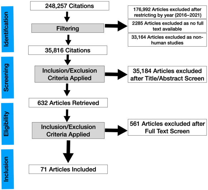
In vivo confocal microscopy (IVCM) imaging is increasingly popular in ocular surface disease diagnosis and management. We conducted a systematic review to update the use of IVCM in the diagnosis and treatment of dry eye and meibomian gland dysfunction (MGD). A literature review was conducted on IVCM studies in MGD, dry eye disease, systemic disease causing dry eye, dry eye in glaucoma patients, contact lens-associated ocular conditions, graft-versus-host disease, and Sjogren's syndrome-related dry eye. The articles were identified through PubMed and a total number of 63 eligible publications were analyzed in detail. All primary research studies on confocal microscopy on dry eye and related conditions from 2017 onwards were included. The reports were reviewed for their contribution to the existing literature as well as potential biases and drawbacks. Despite limitations such as small field of view, lack of population-based norms, and lack of standardization of image acquisition, interpretation, and quantification, IVCM is useful as a complementary technique for clinical diagnosis in various ocular surface disorders related to dry eye. With advances in hardware and software in the near future, it has the potential for further practical impact.

摘要

体内共聚焦显微镜检查(IVCM)成像在眼表疾病的诊断和管理中越来越受欢迎。我们进行了一项系统综述,以更新IVCM在干眼和睑板腺功能障碍(MGD)诊断和治疗中的应用。对关于MGD、干眼疾病、导致干眼的全身性疾病、青光眼患者的干眼、与隐形眼镜相关的眼部疾病、移植物抗宿主病以及干燥综合征相关干眼的IVCM研究进行了文献综述。通过PubMed检索文章,共详细分析了63篇符合条件的出版物。纳入了2017年以来所有关于干眼及相关病症共聚焦显微镜检查的主要研究。对这些报告对现有文献的贡献以及潜在的偏倚和缺点进行了审查。尽管存在诸如视野小、缺乏基于人群的标准以及图像采集、解读和量化缺乏标准化等局限性,但IVCM作为一种辅助技术,在各种与干眼相关的眼表疾病的临床诊断中是有用的。随着硬件和软件在不久的将来取得进展,它有可能产生更大的实际影响。

https://cdn.ncbi.nlm.nih.gov/pmc/blobs/a3ea/9099706/fbed8492ae1f/jcm-11-02349-g001.jpg

相似文献

1
In Vivo Confocal Microscopy in Different Types of Dry Eye and Meibomian Gland Dysfunction.不同类型干眼和睑板腺功能障碍的体内共聚焦显微镜检查
J Clin Med. 2022 Apr 22;11(9):2349. doi: 10.3390/jcm11092349.
2
[A new classification for meibomian gland diseases with in vivo confocal microscopy].[一种用于睑板腺疾病的新型体内共聚焦显微镜分类法]
J Fr Ophtalmol. 2016 Mar;39(3):239-47. doi: 10.1016/j.jfo.2015.07.015. Epub 2016 Feb 16.
3
In vivo confocal microscopy classification in the diagnosis of meibomian gland dysfunction.在活体共聚焦显微镜分类诊断中的睑板腺功能障碍。
Eye (Lond). 2019 May;33(5):754-760. doi: 10.1038/s41433-018-0307-9. Epub 2018 Dec 12.
4
Confocal Microscopy Evaluation of Meibomian Gland Dysfunction in Dry Eye Patients with Different Symptoms.不同症状干眼患者睑板腺功能障碍的共聚焦显微镜评估
Chin Med J (Engl). 2016 Nov 5;129(21):2617-2622. doi: 10.4103/0366-6999.192782.
5
In vivo detection of clinically non-apparent ocular surface inflammation in patients with meibomian gland dysfunction-associated refractory dry eye symptoms: a pilot study.睑板腺功能障碍相关难治性干眼症状患者临床隐匿性眼表炎症的体内检测:一项初步研究。
Eye (Lond). 2015 Aug;29(8):1099-110. doi: 10.1038/eye.2015.103. Epub 2015 Jun 19.
6
In vivo confocal microscopy evaluation of ocular and cutaneous alterations in patients with rosacea.酒渣鼻患者眼部和皮肤改变的体内共聚焦显微镜评估
Br J Ophthalmol. 2017 Mar;101(3):268-274. doi: 10.1136/bjophthalmol-2015-308110. Epub 2016 May 24.
7
Clinical Analysis: Aqueous-Deficient and Meibomian Gland Dysfunction in Patients With Primary Sjogren's Syndrome.临床分析:原发性干燥综合征患者的水液缺乏性干眼和睑板腺功能障碍
Front Med (Lausanne). 2019 Dec 10;6:291. doi: 10.3389/fmed.2019.00291. eCollection 2019.
8
[In vivo confocal microscopy evaluation of meibomian glands in meibomian gland dysfunction patients].睑板腺功能障碍患者睑板腺的体内共聚焦显微镜评估
Zhonghua Yan Ke Za Zhi. 2016 Sep 11;52(9):649-56. doi: 10.3760/cma.j.issn.0412-4081.2016.09.004.
9
Meibomian gland dysfunction and primary Sjögren's syndrome dry eye: a protocol for systematic review and meta-analysis.睑板腺功能障碍与原发性干燥综合征干眼:系统评价和荟萃分析方案。
BMJ Open. 2021 Dec 30;11(12):e048336. doi: 10.1136/bmjopen-2020-048336.
10
Correlation Study Between Abnormal Morphology of Meibomian Glands and Meibum in Patients With Dry Eye Disease Under Confocal Microscopy.干眼患者睑板腺及睑脂形态异常在共聚焦显微镜下的相关性研究
Front Med (Lausanne). 2022 Jan 12;8:793338. doi: 10.3389/fmed.2021.793338. eCollection 2021.

引用本文的文献

1
In Vivo Confocal Microscopy for Automated Detection of Meibomian Gland Dysfunction: A Study Based on Deep Convolutional Neural Networks.用于睑板腺功能障碍自动检测的体内共聚焦显微镜检查:基于深度卷积神经网络的研究
J Imaging Inform Med. 2025 Jan 27. doi: 10.1007/s10278-024-01174-y.
2
A review of the application of in-vivo confocal microscopy on conjunctival diseases.共聚焦显微镜在结膜疾病中的应用综述
Eye Vis (Lond). 2024 Nov 1;11(1):43. doi: 10.1186/s40662-024-00409-x.
3
Redefining our vision: an updated guide to the ocular immune system.

本文引用的文献

1
Diabetic corneal neuropathy as a surrogate marker for diabetic peripheral neuropathy.糖尿病性角膜神经病变作为糖尿病周围神经病变的替代标志物。
Neural Regen Res. 2022 Oct;17(10):2172-2178. doi: 10.4103/1673-5374.327364.
2
Wide-field mosaics of the corneal subbasal nerve plexus in Parkinson's disease using in vivo confocal microscopy.应用活体共聚焦显微镜观察帕金森病患者角膜基底部神经丛的广角镶嵌图像。
Sci Data. 2021 Nov 26;8(1):306. doi: 10.1038/s41597-021-01087-3.
3
In vivo confocal microscopy assessment of meibomian glands microstructure in patients with Graves' orbitopathy.
重新定义我们的视野:眼部免疫系统更新指南。
Nat Rev Immunol. 2024 Dec;24(12):896-911. doi: 10.1038/s41577-024-01064-y. Epub 2024 Aug 30.
4
Quality of Life and Symptomatology in Neuropathic Corneal Pain in Comparison With Dry Eye Syndrome.与干眼综合征相比,神经性角膜疼痛患者的生活质量和症状学表现
Cornea. 2024 Aug 20. doi: 10.1097/ICO.0000000000003674.
5
Confocal Microscopy of the Cornea in Aqueous-Deficient Dry Eye Disease-A Literature Review.水液缺乏型干眼疾病中角膜的共聚焦显微镜检查——文献综述
Diagnostics (Basel). 2024 Jul 26;14(15):1613. doi: 10.3390/diagnostics14151613.
6
Artificial-Intelligence-Enhanced Analysis of In Vivo Confocal Microscopy in Corneal Diseases: A Review.人工智能增强的角膜疾病体内共聚焦显微镜分析:综述
Diagnostics (Basel). 2024 Mar 26;14(7):694. doi: 10.3390/diagnostics14070694.
7
Inflammation mechanism and anti-inflammatory therapy of dry eye.干眼的炎症机制与抗炎治疗
Front Med (Lausanne). 2024 Feb 14;11:1307682. doi: 10.3389/fmed.2024.1307682. eCollection 2024.
8
International survey on dry eye diagnosis by experts.专家关于干眼诊断的国际调查。
Heliyon. 2023 Jun 7;9(6):e16995. doi: 10.1016/j.heliyon.2023.e16995. eCollection 2023 Jun.
9
Role of confocal microscopy in dry eye disease and eye pain.共聚焦显微镜在干眼和眼痛中的作用。
Indian J Ophthalmol. 2023 Apr;71(4):1099-1104. doi: 10.4103/IJO.IJO_3013_22.
10
Enhancing Clinical Decision-Making in Complex Corneal Disorders: The Role of In-Vivo Confocal Microscopy.增强复杂角膜疾病的临床决策:活体共聚焦显微镜的作用
Life (Basel). 2023 Mar 2;13(3):679. doi: 10.3390/life13030679.
Graves 眼病患者睑板腺微观结构的活体共聚焦显微镜评估。
BMC Ophthalmol. 2021 Jun 19;21(1):261. doi: 10.1186/s12886-021-02024-z.
4
Quantification of Increased Corneal Subbasal Nerve Tortuosity in Dry Eye Disease and Its Correlation With Clinical Parameters.定量分析干燥性眼病患者角膜基底部神经扭曲度增加及其与临床参数的相关性。
Transl Vis Sci Technol. 2021 May 3;10(6):26. doi: 10.1167/tvst.10.6.26.
5
Corneal nerve structure in patients with primary Sjögren's syndrome in China.中国原发性干燥综合征患者的角膜神经结构。
BMC Ophthalmol. 2021 May 12;21(1):211. doi: 10.1186/s12886-021-01967-7.
6
In vivo confocal microscopy study of corneal nerve alterations in children and youths with Type 1 diabetes.1 型糖尿病患儿和青少年角膜神经改变的活体共聚焦显微镜研究。
Pediatr Diabetes. 2021 Aug;22(5):780-786. doi: 10.1111/pedi.13219. Epub 2021 May 11.
7
Confocal Microscopic Evaluation of Corneal Dendritic Cell Density and Subbasal Nerve Parameters in Dry Eye Patients: A Systematic Review and Meta-analysis.干眼患者角膜树突状细胞密度和基底神经参数的共聚焦显微镜评估:系统评价与Meta分析
Front Med (Lausanne). 2021 Apr 7;8:578233. doi: 10.3389/fmed.2021.578233. eCollection 2021.
8
The Association between Meibomian Gland Atrophy and Corneal Subbasal Nerve Loss in Patients with Chronic Ocular Graft-versus-host Disease.慢性眼部移植物抗宿主病患者的睑板腺萎缩与角膜基底神经丢失的相关性。
Curr Eye Res. 2021 Jun;46(6):796-801. doi: 10.1080/02713683.2020.1846754. Epub 2021 Jan 11.
9
Diabetic Corneal Neuropathy.糖尿病性角膜神经病变
J Clin Med. 2020 Dec 6;9(12):3956. doi: 10.3390/jcm9123956.
10
Multiwavelength confocal laser scanning microscopy of the cornea.角膜的多波长共聚焦激光扫描显微镜检查
Biomed Opt Express. 2020 Sep 18;11(10):5689-5700. doi: 10.1364/BOE.397615. eCollection 2020 Oct 1.