Matsuo-Hagiyama Chisato, Watanabe Yoshiyuki, Tanaka Hisashi, Takahashi Hiroto, Arisawa Atsuko, Yoshioka Eri, Nabatame Shin, Nakano Sayaka, Tomiyama Noriyuki
Diagnostic and Interventional Radiology, Osaka University Graduate School of Medicine.
Department of Pediatrics, Osaka University Graduate School of Medicine.
Magn Reson Med Sci. 2017 Jul 10;16(3):209-216. doi: 10.2463/mrms.mp.2016-0045. Epub 2016 Oct 31.
Silent magnetic resonance imaging (MRI) scans produce reduced acoustic noise and are considered more gentle for sedated children. The aim of this study was to compare the validity of T- (TW) and T-weighted (TW) silent sequences for myelination assessment in children with conventional spin-echo sequences.
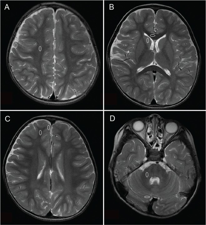
A total of 30 children (21 boys, 9 girls; age range: 1-83 months, mean age: 35.5 months, median age: 28.5 months) were examined using both silent and spin-echo sequences. Acoustic noise levels were analyzed and compared. The degree of myelination was qualitatively assessed via consensus, and TW and TW signal intensities were quantitatively measured by percent contrast.
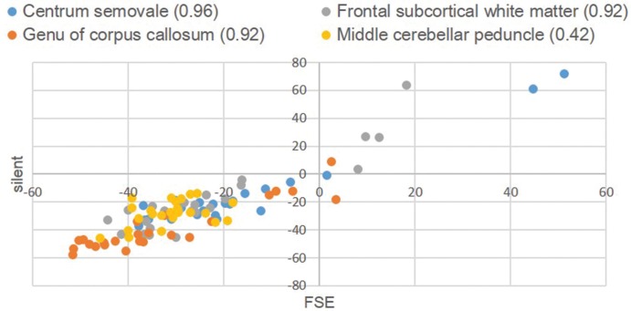
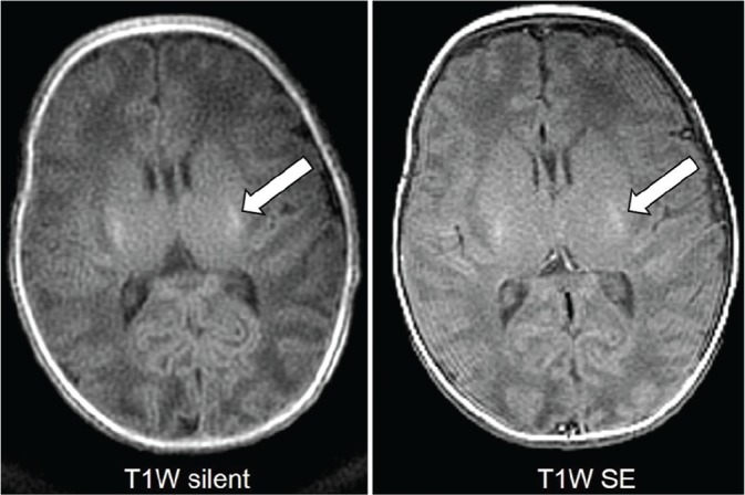
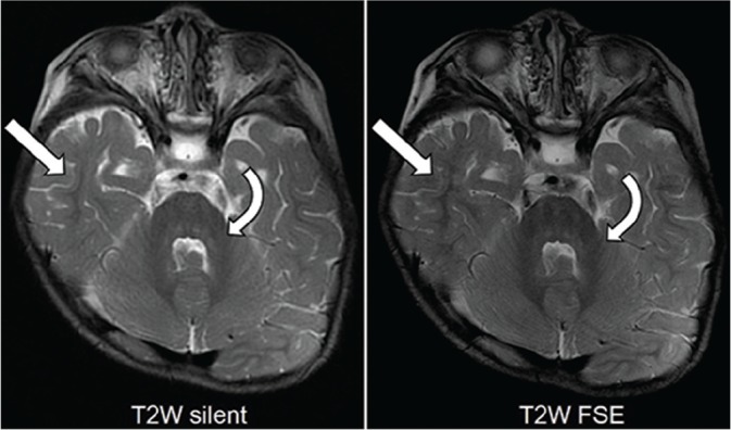
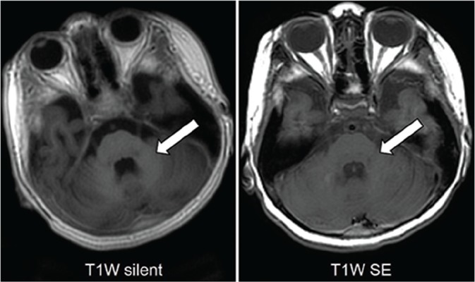
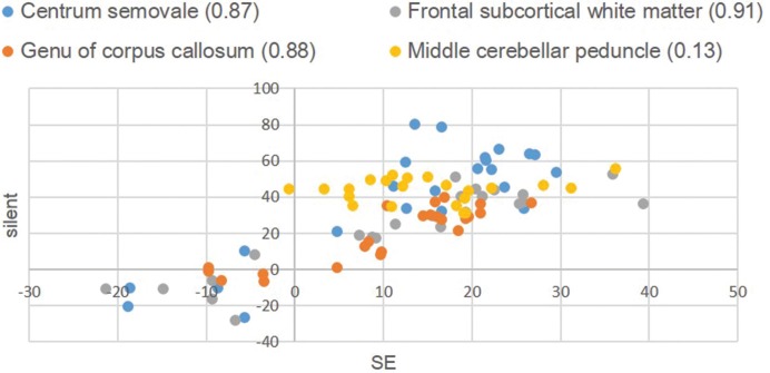
Acoustic noise levels were significantly lower during silent sequences than during conventional sequences (P < 0.0001 for both TW and TW). Inter-method comparison indicated overall good to excellent agreement (TW and TW images, κ = 0.76 and 0.80, respectively); however, agreement was poor for cerebellar myelination on TW images (κ = 0.14). The percent contrast of silent and conventional MRI sequences had a strong correlation (TW, correlation coefficient [CC] = 0.76; TW excluding the middle cerebellar peduncle, CC = 0.82; TW, CC = 0.91).
For brain MRI, silent sequences significantly reduced acoustic noise and provided diagnostic image quality for myelination evaluations; however, the two methods differed with respect to cerebellar delineation on TW sequences.
静音磁共振成像(MRI)扫描产生的声学噪声较低,被认为对接受镇静的儿童更为温和。本研究的目的是比较T-(TW)加权和T加权静音序列与传统自旋回波序列在评估儿童髓鞘形成方面的有效性。
共有30名儿童(21名男孩,9名女孩;年龄范围:1 - 83个月,平均年龄:35.5个月,中位年龄:28.5个月)接受了静音和自旋回波序列检查。分析并比较了声学噪声水平。通过共识定性评估髓鞘形成程度,并通过对比百分比定量测量TW和TW信号强度。
静音序列期间的声学噪声水平明显低于传统序列(TW和TW均P < 0.0001)。方法间比较表明总体一致性良好至优秀(TW和TW图像,κ分别为0.76和0.80);然而,TW图像上小脑髓鞘形成的一致性较差(κ = 0.14)。静音和传统MRI序列的对比百分比具有很强的相关性(TW,相关系数[CC] = 0.76;排除小脑中脚的TW,CC = 0.82;TW,CC = 0.91)。
对于脑部MRI,静音序列显著降低了声学噪声,并为髓鞘形成评估提供了诊断图像质量;然而,两种方法在TW序列上的小脑描绘方面存在差异。