Hegazy Mohamed A A, Cho Min Hyoung, Lee Soo Yeol
Department of Biomedical Engineering, Kyung Hee University, Yongin-Si, Gyeonggi-do, 446-701, South Korea.
Biomed Eng Online. 2016 Nov 4;15(1):119. doi: 10.1186/s12938-016-0240-8.
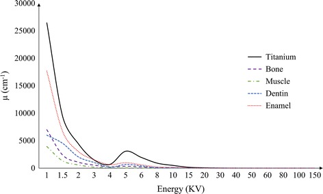
Metal artifacts appearing as streaks and shadows often compromise readability of computed tomography (CT) images. Particularly in a dental CT in which high resolution imaging is crucial for precise preparation of dental implants or orthodontic devices, reduction of metal artifacts is very important. However, metal artifact reduction algorithms developed for a general medical CT may not work well in a dental CT since teeth themselves also have high attenuation coefficients.
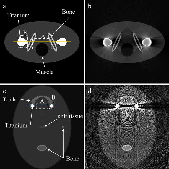
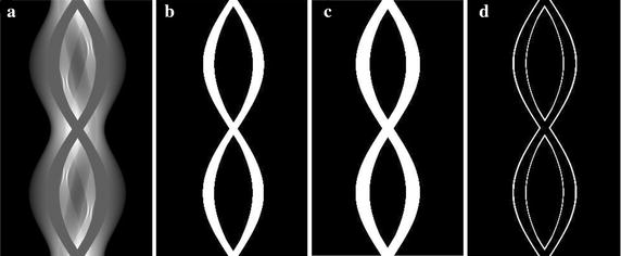
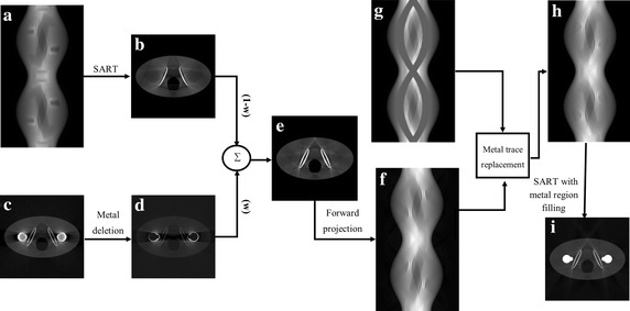
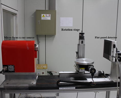
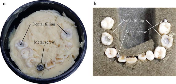
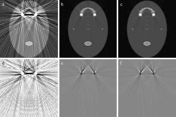
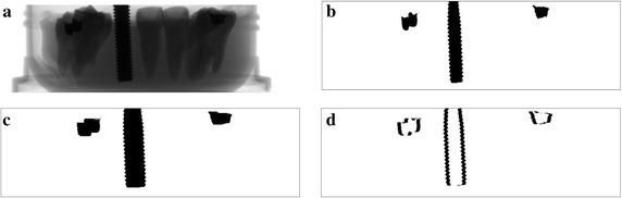
To reduce metal artifacts in dental CT images, we made prior images by weighted summation of two images: one, a streak-reduced image reconstructed from the metal-region-modified projection data, and the other a metal-free image reconstructed from the original projection data followed by metal region deletion. To make the streak-reduced image, we precisely segmented the metal region based on adaptive local thresholding, and then, we modified the metal region on the projection data using linear interpolation. We made forward projection of the prior image to make the prior projection data. We replaced the pixel values at the metal region in the original projection data with the ones taken from the prior projection data, and then, we finally reconstructed images from the replaced projection data. To validate the proposed method, we made computational simulations and also we made experiments on teeth phantoms using a micro-CT. We compared the results with the ones obtained by the fusion prior-based metal artifact reduction (FP-MAR) method.
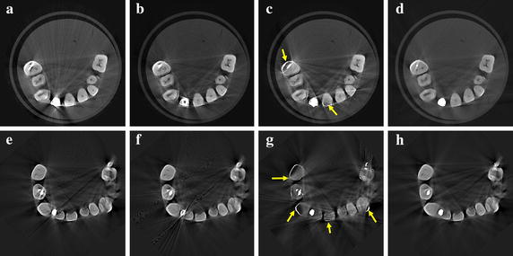
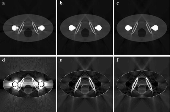
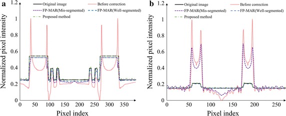
In the simulation studies using a bilateral prostheses phantom and a dental phantom, the proposed method showed a performance similar to the FP-MAR method in terms of the edge profile and the structural similarity index when an optimal global threshold was chosen for the FP-MAR method. In the imaging studies of teeth phantoms, the proposed method showed a better performance than the FP-MAR method in reducing the streak artifacts without introducing any contrast anomaly.
The simulation and experimental imaging studies suggest that the proposed method can be used for reducing metal artifacts in dental CT images.
表现为条纹和阴影的金属伪影常常会影响计算机断层扫描(CT)图像的可读性。特别是在牙科CT中,高分辨率成像对于精确制备牙种植体或正畸器械至关重要,减少金属伪影非常重要。然而,为普通医学CT开发的金属伪影减少算法在牙科CT中可能效果不佳,因为牙齿本身也具有高衰减系数。
为了减少牙科CT图像中的金属伪影,我们通过对两幅图像进行加权求和来制作先验图像:一幅是从金属区域修改后的投影数据重建的条纹减少图像,另一幅是从原始投影数据重建并随后删除金属区域的无金属图像。为了制作条纹减少图像,我们基于自适应局部阈值精确分割金属区域,然后使用线性插值对投影数据上的金属区域进行修改。我们对先验图像进行正投影以制作先验投影数据。我们将原始投影数据中金属区域的像素值替换为先验投影数据中的像素值,然后最终从替换后的投影数据重建图像。为了验证所提出的方法,我们进行了计算模拟,并使用微型CT对牙齿模型进行了实验。我们将结果与基于融合先验的金属伪影减少(FP-MAR)方法获得的结果进行了比较。
在使用双侧假体模型和牙科模型的模拟研究中,当为FP-MAR方法选择最佳全局阈值时,所提出的方法在边缘轮廓和结构相似性指数方面表现出与FP-MAR方法相似的性能。在牙齿模型的成像研究中,所提出的方法在减少条纹伪影方面表现优于FP-MAR方法,且未引入任何对比度异常。
模拟和实验成像研究表明,所提出的方法可用于减少牙科CT图像中的金属伪影。