Molino Yves, Jabès Françoise, Bonnet Amandine, Gaudin Nicolas, Bernard Anne, Benech Philippe, Khrestchatisky Michel
Vect-Horus SAS, Faculté de Médecine - Secteur Nord, 51 Bd Pierre Dramard, 13344, Marseille Cedex 15, France.
Aix Marseille Univ, CNRS, NICN, Marseille, France.
J Neuroinflammation. 2016 Nov 10;13(1):290. doi: 10.1186/s12974-016-0749-6.
The heterogeneity of endothelial cell types underlies their remarkable ability to sub-specialize and provide specific requirements for a given vascular bed. Here, we compared rat microvascular endothelial cells (MECs) derived from the brain and spinal cord in both basal and inflammatory conditions.
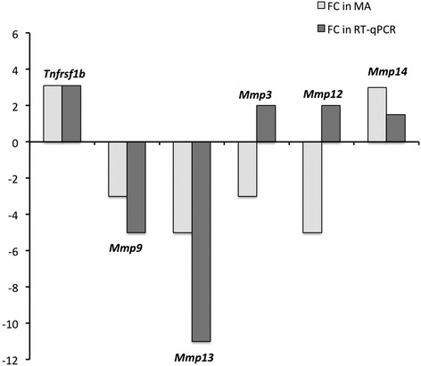
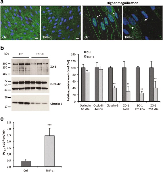
We used whole rat genome microarrays to compare, at different time points, basal and TNF-α-induced gene expression of rat MECs from in vitro models of the blood-brain barrier (BBB) and blood-spinal cord barrier (BSCB). Validation at both messenger RNA (mRNA) and protein levels was performed on freshly extracted microvessels (MVs) from the brain and spinal cord (BMVs and SCMVs, respectively), as these were considered the closest in vivo tissues to cultured MECs.
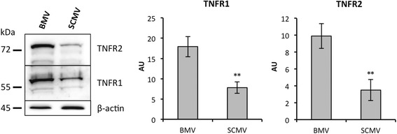
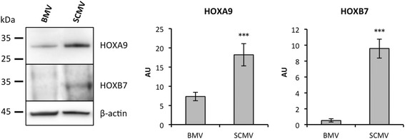
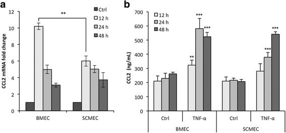
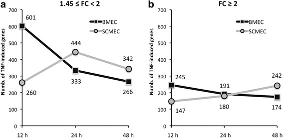
Most of the genes encoding adhesion/tight junction molecules and known endothelial markers were similarly expressed in brain and spinal cord MECs (BMECs and SCMECs, respectively). However, one striking finding was the higher expression of several Hox genes, which encode transcription factors involved in positional identity. The differential expression of Hoxa9 and Hoxb7 at the mRNA levels as well as protein levels was confirmed in BMVs and SCMVs. Although the TNF-α response was in general higher in BMECs than in SCMECs at 12 h, the opposite was observed at 48 h. Furthermore, we found that expression of Tnfrsf1a and Tnfrsf1b encoding the TNF receptor super-family member 1a/TNFR1 and 1b/TNFR2, respectively, were constitutively higher in BMVs compared to SCMVs. However, only Tnfrsf1b was induced in SCMECs in response to TNF-α at 24 and 48 h.
Our results support a role for HOX members in defining the positional identities of MECs in vivo. Our data also suggest that the delayed transcriptional activation upon TNF-α treatment in SCMECs results from the requirement of the TNF-induced expression of Tnfrsf1b. In contrast, its high basal expression in BMECs might be sufficient to confer an immediate and efficient TNF-α response.
内皮细胞类型的异质性是其显著的亚专业化能力的基础,并为特定的血管床提供特定需求。在此,我们比较了在基础状态和炎症状态下源自大脑和脊髓的大鼠微血管内皮细胞(MECs)。
我们使用全大鼠基因组微阵列,在不同时间点比较来自血脑屏障(BBB)和血脊髓屏障(BSCB)体外模型的大鼠MECs的基础基因表达和肿瘤坏死因子-α(TNF-α)诱导的基因表达。在分别从大脑和脊髓新鲜提取的微血管(MVs)(分别为脑微血管和脊髓微血管)上进行信使核糖核酸(mRNA)和蛋白质水平的验证,因为这些被认为是与培养的MECs最接近的体内组织。
大多数编码黏附/紧密连接分子和已知内皮标志物的基因在脑和脊髓MECs(分别为BMECs和SCMECs)中表达相似。然而,一个显著的发现是几个Hox基因的表达较高,这些基因编码参与位置身份的转录因子。Hoxa9和Hoxb7在mRNA水平以及蛋白质水平的差异表达在脑微血管和脊髓微血管中得到证实。虽然在12小时时,BMECs中TNF-α反应总体上高于SCMECs,但在48小时时观察到相反的情况。此外,我们发现分别编码TNF受体超家族成员1a/TNFR1和1b/TNFR2的Tnfrsf1a和Tnfrsf1b的表达在脑微血管中相比脊髓微血管中组成性地更高。然而,仅Tnfrsf1b在SCMECs中在24和48小时时对TNF-α有诱导反应。
我们的结果支持HOX成员在体内定义MECs位置身份中的作用。我们的数据还表明,SCMECs中TNF-α处理后转录激活延迟是由于TNF诱导的Tnfrsf1b表达的需求。相反,其在BMECs中的高基础表达可能足以赋予即时且有效的TNF-α反应。