Olvera-García Gustavo, Aguilar-García Tania, Gutiérrez-Jasso Fany, Imaz-Rosshandler Iván, Rangel-Escareño Claudia, Orozco Lorena, Aguilar-Delfín Irma, Vázquez-Pérez Joel A, Zúñiga Joaquín, Pérez-Patrigeon Santiago, Espinosa Enrique
Department of Research in Immunology, Instituto Nacional de Enfermedades Respiratorias Ismael Cosío Villegas, Calzada de Tlalpan 4502, Mexico City, Mexico.
Computational Genomics Department, Instituto Nacional de Medicina Genómica, Periferico Sur 4809, Mexico City, Mexico.
BMC Genomics. 2016 Nov 22;17(1):956. doi: 10.1186/s12864-016-3308-8.
Human central memory CD4 T cells are characterized by their capacity of proliferation and differentiation into effector memory CD4 T cells. Homeostasis of central memory CD4 T cells is considered a key factor sustaining the asymptomatic stage of Human Immunodeficiency Virus type 1 (HIV-1) infection, while progression to acquired immunodeficiency syndrome is imputed to central memory CD4 T cells homeostatic failure. We investigated if central memory CD4 T cells from patients with HIV-1 infection have a gene expression profile impeding proliferation and survival, despite their activated state.
Using gene expression microarrays, we analyzed mRNA expression patterns in naive, central memory, and effector memory CD4 T cells from healthy controls, and naive and central memory CD4 T cells from patients with HIV-1 infection. Differentially expressed genes, defined by Log Fold Change (FC) ≥ |0.5| and Log (odds) > 0, were used in pathway enrichment analyses.
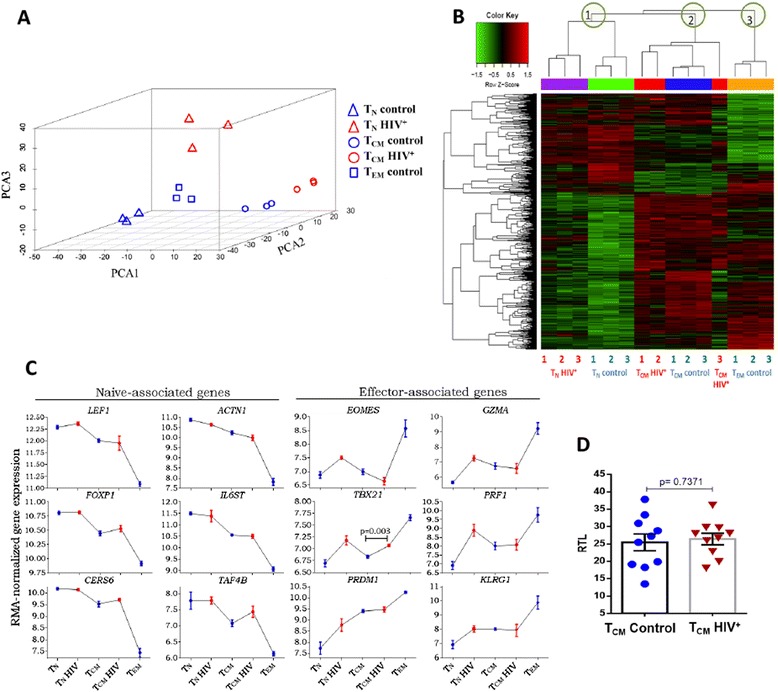
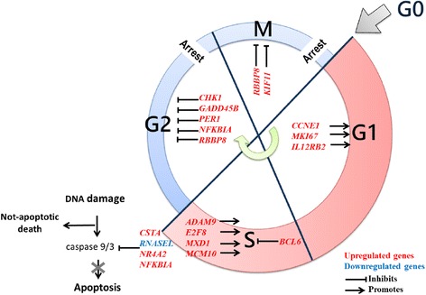
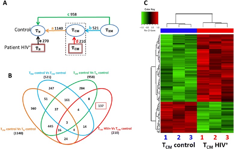
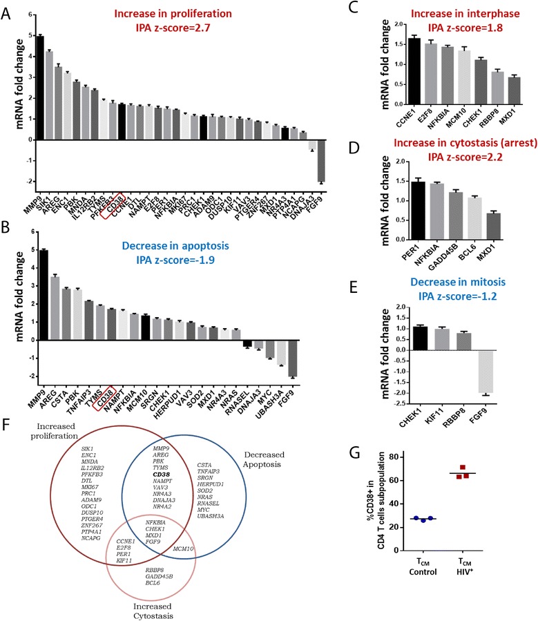
Central memory CD4 T cells from patients and controls showed comparable expression of differentiation-related genes, ruling out an effector-like differentiation of central memory CD4 T cells in HIV infection. However, 210 genes were differentially expressed in central memory CD4 T cells from patients compared with those from controls. Expression of 75 of these genes was validated by semi quantitative RT-PCR, and independently reproduced enrichment results from this gene expression signature. The results of functional enrichment analysis indicated movement to cell cycle phases G1 and S (increased CCNE1, MKI67, IL12RB2, ADAM9, decreased FGF9, etc.), but also arrest in G2/M (increased CHK1, RBBP8, KIF11, etc.). Unexpectedly, the results also suggested decreased apoptosis (increased CSTA, NFKBIA, decreased RNASEL, etc.). Results also suggested increased IL-1β, IFN-γ, TNF, and RANTES (CCR5) activity upstream of the central memory CD4 T cells signature, consistent with the demonstrated milieu in HIV infection.
Our findings support a model where progressive loss of central memory CD4 T cells in chronic HIV-1 infection is driven by increased cell cycle entry followed by mitotic arrest, leading to a non-apoptotic death pathway without actual proliferation, possibly contributing to increased turnover.
人类中枢记忆性CD4 T细胞的特征在于其增殖以及分化为效应记忆性CD4 T细胞的能力。中枢记忆性CD4 T细胞的稳态被认为是维持1型人类免疫缺陷病毒(HIV-1)感染无症状期的关键因素,而获得性免疫缺陷综合征的进展则归因于中枢记忆性CD4 T细胞的稳态失衡。我们研究了HIV-1感染患者的中枢记忆性CD4 T细胞是否具有阻碍增殖和存活的基因表达谱,尽管它们处于激活状态。
使用基因表达微阵列,我们分析了来自健康对照的初始、中枢记忆和效应记忆CD4 T细胞以及来自HIV-1感染患者的初始和中枢记忆CD4 T细胞中的mRNA表达模式。通过对数倍变化(FC)≥|0.5|和对数(优势)>0定义的差异表达基因用于通路富集分析。
患者和对照的中枢记忆性CD4 T细胞显示出分化相关基因的可比表达,排除了HIV感染中枢记忆性CD4 T细胞的效应样分化。然而,与对照相比,患者的中枢记忆性CD4 T细胞中有210个基因差异表达。这些基因中的75个基因的表达通过半定量RT-PCR得到验证,并独立重现了该基因表达特征的富集结果。功能富集分析结果表明细胞周期进入G1和S期(CCNE1、MKI67、IL12RB2、ADAM9增加,FGF9减少等),但也停滞在G2/M期(CHK1、RBBP8、KIF11增加等)。出乎意料的是,结果还表明凋亡减少(CSTA、NFKBIA增加,RNASEL减少等)。结果还表明中枢记忆性CD4 T细胞特征上游的IL-1β、IFN-γ、TNF和RANTES(CCR5)活性增加,与HIV感染中已证实的环境一致。
我们的研究结果支持一种模型,即慢性HIV-1感染中中枢记忆性CD4 T细胞的逐渐丧失是由细胞周期进入增加随后有丝分裂停滞驱动的,导致无实际增殖的非凋亡死亡途径,可能导致周转率增加。