Paul Colin D, Mistriotis Panagiotis, Konstantopoulos Konstantinos
Department of Chemical and Biomolecular Engineering and the Institute for NanoBioTechnology, Johns Hopkins University, 3400 North Charles Street, Baltimore, Maryland 21218, USA.
Department of Biomedical Engineering, Johns Hopkins University, 3400 North Charles Street, Baltimore, Maryland 21218, USA.
Nat Rev Cancer. 2017 Feb;17(2):131-140. doi: 10.1038/nrc.2016.123. Epub 2016 Dec 2.
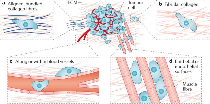
Time-lapse, deep-tissue imaging made possible by advances in intravital microscopy has demonstrated the importance of tumour cell migration through confining tracks in vivo. These tracks may either be endogenous features of tissues or be created by tumour or tumour-associated cells. Importantly, migration mechanisms through confining microenvironments are not predicted by 2D migration assays. Engineered in vitro models have been used to delineate the mechanisms of cell motility through confining spaces encountered in vivo. Understanding cancer cell locomotion through physiologically relevant confining tracks could be useful in developing therapeutic strategies to combat metastasis.
活体显微镜技术的进步使延时、深部组织成像成为可能,这已证明肿瘤细胞在体内通过狭窄通道迁移的重要性。这些通道可能是组织的内源性特征,也可能是由肿瘤或肿瘤相关细胞形成的。重要的是,二维迁移试验无法预测通过狭窄微环境的迁移机制。工程化的体外模型已被用于描绘细胞通过体内遇到的狭窄空间的运动机制。了解癌细胞通过生理相关的狭窄通道的运动,可能有助于制定对抗转移的治疗策略。