• 文献检索
  • 文档翻译
  • 深度研究
  • 学术资讯
  • Suppr Zotero 插件Zotero 插件
  • 邀请有礼
  • 套餐&价格
  • 历史记录
应用&插件
Suppr Zotero 插件Zotero 插件浏览器插件Mac 客户端Windows 客户端微信小程序
定价
高级版会员购买积分包购买API积分包
服务
文献检索文档翻译深度研究API 文档MCP 服务
关于我们
关于 Suppr公司介绍联系我们用户协议隐私条款
关注我们

Suppr 超能文献

核心技术专利:CN118964589B侵权必究
粤ICP备2023148730 号-1Suppr @ 2026

文献检索

告别复杂PubMed语法,用中文像聊天一样搜索,搜遍4000万医学文献。AI智能推荐,让科研检索更轻松。

立即免费搜索

文件翻译

保留排版,准确专业,支持PDF/Word/PPT等文件格式,支持 12+语言互译。

免费翻译文档

深度研究

AI帮你快速写综述,25分钟生成高质量综述,智能提取关键信息,辅助科研写作。

立即免费体验

准包膜甲型肝炎病毒的胆汁分泌

Biliary Secretion of Quasi-Enveloped Human Hepatitis A Virus.

作者信息

Hirai-Yuki Asuka, Hensley Lucinda, Whitmire Jason K, Lemon Stanley M

机构信息

Lineberger Comprehensive Cancer Center, the University of North Carolina at Chapel Hill, Chapel Hill, North Carolina, USA.

Department of Microbiology & Immunology, the University of North Carolina at Chapel Hill, Chapel Hill, North Carolina, USA.

出版信息

mBio. 2016 Dec 6;7(6):e01998-16. doi: 10.1128/mBio.01998-16.

DOI:10.1128/mBio.01998-16
PMID:27923925
原文链接:https://pmc.ncbi.nlm.nih.gov/articles/PMC5142623/
Abstract

UNLABELLED

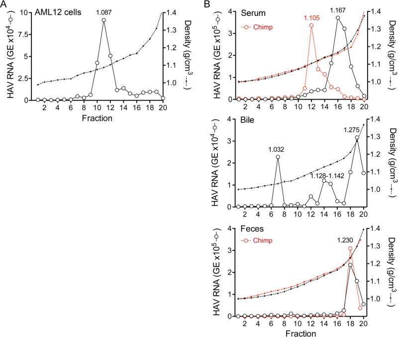
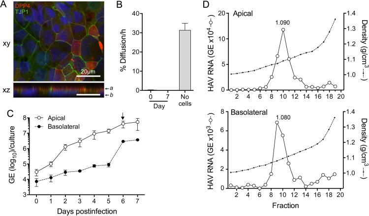
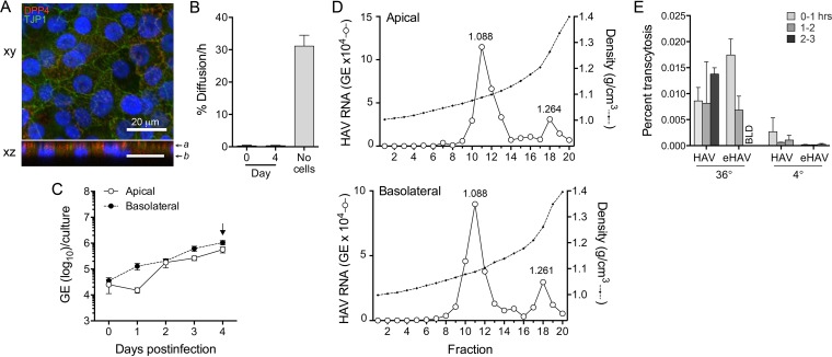
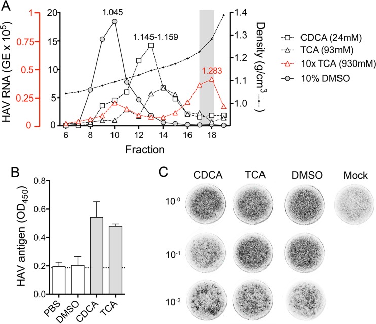
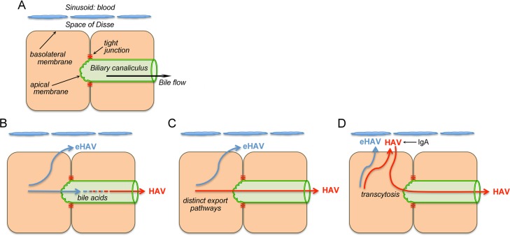
Hepatitis A virus (HAV) is an unusual picornavirus that is released from cells cloaked in host-derived membranes. These quasi-enveloped virions (eHAV) are the only particle type circulating in blood during infection, whereas only nonenveloped virions are shed in feces. The reason for this is uncertain. Hepatocytes, the only cell type known to support HAV replication in vivo, are highly polarized epithelial cells with basolateral membranes facing onto hepatic (blood) sinusoids and apical membranes abutting biliary canaliculi from which bile is secreted to the gut. To assess whether eHAV and nonenveloped virus egress from cells via vectorially distinct pathways, we studied infected polarized cultures of Caco-2 and HepG2-N6 cells. Most (>99%) progeny virions were released apically from Caco-2 cells, whereas basolateral (64%) versus apical (36%) release was more balanced with HepG2-N6 cells. Both apically and basolaterally released virions were predominantly enveloped, with no suggestion of differential vectorial release of eHAV versus naked virions. Basolateral to apical transcytosis of either particle type was minimal (<0.02%/h) in HepG2-N6 cells, arguing against this as a mechanism for differences in membrane envelopment of serum versus fecal virus. High concentrations of human bile acids converted eHAV to nonenveloped virions, whereas virus present in bile from HAV-infected Ifnar1 Ifngr1 and Mavs mice banded over a range of densities extending from that of eHAV to that of nonenveloped virions. We conclude that nonenveloped virions shed in feces are derived from eHAV released across the canalicular membrane and stripped of membranes by the detergent action of bile acids within the proximal biliary canaliculus.

IMPORTANCE

HAV is a hepatotropic, fecally/orally transmitted picornavirus that can cause severe hepatitis in humans. Recent work reveals that it has an unusual life cycle. Virus is found in cell culture supernatant fluids in two mature, infectious forms: one wrapped in membranes (quasi-enveloped) and another that is nonenveloped. Membrane-wrapped virions circulate in blood during acute infection and are resistant to neutralizing antibodies, likely facilitating HAV dissemination within the liver. On the other hand, virus shed in feces is nonenveloped and highly stable, facilitating epidemic spread and transmission to naive hosts. Factors controlling the biogenesis of these two distinct forms of the virus in infected humans are not understood. Here we characterize vectorial release of quasi-enveloped virions from polarized epithelial cell cultures and provide evidence that bile acids strip membranes from eHAV following its secretion into the biliary tract. These results enhance our understanding of the life cycle of this unusual picornavirus.

摘要
https://cdn.ncbi.nlm.nih.gov/pmc/blobs/35cf/5142623/f9aecceeb642/mbo0061630890005.jpg
https://cdn.ncbi.nlm.nih.gov/pmc/blobs/35cf/5142623/8d14caf123ee/mbo0061630890001.jpg
https://cdn.ncbi.nlm.nih.gov/pmc/blobs/35cf/5142623/f513474836ea/mbo0061630890002.jpg
https://cdn.ncbi.nlm.nih.gov/pmc/blobs/35cf/5142623/488bfcdd72d5/mbo0061630890003.jpg
https://cdn.ncbi.nlm.nih.gov/pmc/blobs/35cf/5142623/439d68060bf2/mbo0061630890004.jpg
https://cdn.ncbi.nlm.nih.gov/pmc/blobs/35cf/5142623/f9aecceeb642/mbo0061630890005.jpg

未标记

甲型肝炎病毒(HAV)是一种不同寻常的小核糖核酸病毒,它从包裹着宿主来源膜的细胞中释放出来。这些准包膜病毒粒子(eHAV)是感染期间血液中循环的唯一粒子类型,而只有无包膜病毒粒子从粪便中排出。其原因尚不确定。肝细胞是已知在体内支持HAV复制的唯一细胞类型,是高度极化的上皮细胞,其基底外侧膜面向肝(血)窦,顶端膜邻接胆小管,胆汁从胆小管分泌到肠道。为了评估eHAV和无包膜病毒是否通过不同的向量途径从细胞中释放出来,我们研究了Caco-2和HepG2-N6细胞的感染极化培养物。大多数(>99%)子代病毒粒子从Caco-2细胞顶端释放,而HepG2-N6细胞的基底外侧(64%)与顶端(36%)释放更为平衡。顶端和基底外侧释放的病毒粒子主要是包膜的,没有迹象表明eHAV与裸露病毒粒子有不同的向量释放。在HepG2-N6细胞中,两种粒子类型从基底外侧到顶端的跨细胞转运极少(<0.02%/小时),这表明这不是血清病毒与粪便病毒膜包裹差异的机制。高浓度的人胆汁酸将eHAV转化为无包膜病毒粒子,而来自感染HAV的Ifnar1 Ifngr1和Mavs小鼠胆汁中的病毒在一系列密度范围内聚集,从eHAV的密度到无包膜病毒粒子的密度。我们得出结论,粪便中排出的无包膜病毒粒子来源于通过胆小管膜释放的eHAV,并在近端胆小管内被胆汁酸的去污剂作用剥去膜。

重要性

HAV是一种嗜肝、经粪口传播的小核糖核酸病毒,可导致人类严重肝炎。最近的研究表明它有一个不同寻常的生命周期。在细胞培养上清液中发现病毒有两种成熟的感染形式:一种包裹在膜中(准包膜),另一种是无包膜的。膜包裹的病毒粒子在急性感染期间在血液中循环,并且对中和抗体有抗性,这可能促进HAV在肝脏内的传播。另一方面,粪便中排出的病毒是无包膜的且高度稳定,有利于流行传播和传播给未感染的宿主。在受感染的人类中控制这两种不同形式病毒生物发生的因素尚不清楚。在这里,我们描述了准包膜病毒粒子从极化上皮细胞培养物中的向量释放,并提供证据表明胆汁酸在eHAV分泌到胆道后将其膜剥离。这些结果增强了我们对这种不同寻常的小核糖核酸病毒生命周期的理解。

相似文献

1
Biliary Secretion of Quasi-Enveloped Human Hepatitis A Virus.准包膜甲型肝炎病毒的胆汁分泌
mBio. 2016 Dec 6;7(6):e01998-16. doi: 10.1128/mBio.01998-16.
2
TIM1 (HAVCR1) Is Not Essential for Cellular Entry of Either Quasi-enveloped or Naked Hepatitis A Virions.TIM1(HAVCR1)并非甲型肝炎病毒准衣壳或裸病毒进入细胞所必需。
mBio. 2017 Sep 5;8(5):e00969-17. doi: 10.1128/mBio.00969-17.
3
Vectorial Release of Hepatitis E Virus in Polarized Human Hepatocytes.乙型肝炎病毒在极化的人肝细胞中的矢量释放。
J Virol. 2019 Feb 5;93(4). doi: 10.1128/JVI.01207-18. Print 2019 Feb 15.
4
Cellular entry and uncoating of naked and quasi-enveloped human hepatoviruses.裸型和准包膜型人肝炎病毒的细胞进入和脱壳。
Elife. 2019 Feb 25;8:e43983. doi: 10.7554/eLife.43983.
5
Redundant Late Domain Functions of Tandem VP2 YPXL Motifs in Nonlytic Cellular Egress of Quasi-enveloped Hepatitis A Virus.串联 VP2 YPXL 基序的冗余晚期结构域功能在准包膜甲型肝炎病毒非裂解性细胞外溢中的作用。
J Virol. 2018 Nov 12;92(23). doi: 10.1128/JVI.01308-18. Print 2018 Dec 1.
6
Vectorial entry and release of hepatitis A virus in polarized human hepatocytes.甲型肝炎病毒在极化人肝细胞中的向量性进入与释放
J Virol. 2008 Sep;82(17):8733-42. doi: 10.1128/JVI.00219-08. Epub 2008 Jun 25.
7
Naked Viruses That Aren't Always Naked: Quasi-Enveloped Agents of Acute Hepatitis.并非总是赤裸裸的病毒:急性肝炎的准包膜性病原体。
Annu Rev Virol. 2014 Nov;1(1):539-60. doi: 10.1146/annurev-virology-031413-085359. Epub 2014 Jul 3.
8
Protein composition of the hepatitis A virus quasi-envelope.甲型肝炎病毒准衣壳的蛋白组成。
Proc Natl Acad Sci U S A. 2017 Jun 20;114(25):6587-6592. doi: 10.1073/pnas.1619519114. Epub 2017 May 10.
9
Human pDCs preferentially sense enveloped hepatitis A virions.人类浆细胞样树突状细胞优先识别有包膜的甲型肝炎病毒粒子。
J Clin Invest. 2015 Jan;125(1):169-76. doi: 10.1172/JCI77527. Epub 2014 Nov 21.
10
Nonlytic Quasi-Enveloped Hepatovirus Release Is Facilitated by pX Protein Interaction with the E3 Ubiquitin Ligase ITCH.非结构型准衣壳化肝病毒释放由 pX 蛋白与 E3 泛素连接酶 ITCH 的相互作用促进。
J Virol. 2022 Nov 9;96(21):e0119522. doi: 10.1128/jvi.01195-22. Epub 2022 Oct 26.

引用本文的文献

1
The hepatocyte traffic network in the human hepatitis A virus biological cycle from an evolutionary perspective.从进化角度看人类甲型肝炎病毒生物周期中的肝细胞运输网络。
Commun Biol. 2025 Jun 12;8(1):917. doi: 10.1038/s42003-025-08344-w.
2
Hepatitis A and E Viruses Are Important Agents of Acute Severe Hepatitis in Asia: A Narrative Review.甲型和戊型肝炎病毒是亚洲急性重型肝炎的重要病原体:一项叙述性综述
Pathogens. 2025 May 6;14(5):454. doi: 10.3390/pathogens14050454.
3
Enhanced hepatitis E virus infection of polarised hepatocytes in vitro.

本文引用的文献

1
MAVS-dependent host species range and pathogenicity of human hepatitis A virus.人甲型肝炎病毒依赖线粒体抗病毒信号蛋白的宿主物种范围及致病性
Science. 2016 Sep 30;353(6307):1541-1545. doi: 10.1126/science.aaf8325. Epub 2016 Sep 15.
2
Naked Viruses That Aren't Always Naked: Quasi-Enveloped Agents of Acute Hepatitis.并非总是赤裸裸的病毒:急性肝炎的准包膜性病原体。
Annu Rev Virol. 2014 Nov;1(1):539-60. doi: 10.1146/annurev-virology-031413-085359. Epub 2014 Jul 3.
3
Specific IgA Enhances the Transcytosis and Excretion of Hepatitis A Virus.
体外极化肝细胞中戊型肝炎病毒感染增强
Sci Rep. 2025 Mar 4;15(1):7598. doi: 10.1038/s41598-025-92164-9.
4
Preferential apical infection of Caco-2 intestinal cell monolayers by SARS-CoV-2 is associated with damage to cellular barrier integrity: Implications for the pathophysiology of COVID-19.严重急性呼吸综合征冠状病毒2(SARS-CoV-2)对Caco-2肠细胞单层的优先顶端感染与细胞屏障完整性受损有关:对2019冠状病毒病病理生理学的启示。
PLoS One. 2025 Feb 10;20(2):e0313068. doi: 10.1371/journal.pone.0313068. eCollection 2025.
5
A Useful Method to Provide Infectious and Cultivable In Vitro Naked Viral Particles of Hepatitis A Virus.一种提供甲型肝炎病毒传染性和可培养的体外裸病毒颗粒的有用方法。
Viruses. 2024 Aug 26;16(9):1360. doi: 10.3390/v16091360.
6
Biochemical analysis of the host factor activity of ZCCHC14 in hepatitis A virus replication.ZCCHC14 宿主因子在甲型肝炎病毒复制中的生化分析。
J Virol. 2024 Apr 16;98(4):e0005724. doi: 10.1128/jvi.00057-24. Epub 2024 Mar 19.
7
Loxapine inhibits replication of hepatitis A virus in vitro and in vivo by targeting viral protein 2C.洛沙平通过靶向病毒蛋白 2C 抑制甲型肝炎病毒在体外和体内的复制。
PLoS Pathog. 2024 Mar 13;20(3):e1012091. doi: 10.1371/journal.ppat.1012091. eCollection 2024 Mar.
8
Hepatoviruses promote very-long-chain fatty acid and sphingolipid synthesis for viral RNA replication and quasi-enveloped virus release.肝病毒促进长链脂肪酸和神经鞘脂的合成,以进行病毒 RNA 复制和准包膜病毒释放。
Sci Adv. 2023 Oct 20;9(42):eadj4198. doi: 10.1126/sciadv.adj4198.
9
Hepatitis A virus infection.甲型肝炎病毒感染
Nat Rev Dis Primers. 2023 Sep 28;9(1):51. doi: 10.1038/s41572-023-00461-2.
10
Cell entry and release of quasi-enveloped human hepatitis viruses.准包膜人类肝炎病毒的进入和释放。
Nat Rev Microbiol. 2023 Sep;21(9):573-589. doi: 10.1038/s41579-023-00889-z. Epub 2023 Apr 25.
特异性IgA增强甲型肝炎病毒的转胞吞作用和排泄。
Sci Rep. 2016 Feb 25;6:21855. doi: 10.1038/srep21855.
4
Hepatitis A virus and the origins of picornaviruses.甲型肝炎病毒与小核糖核酸病毒的起源
Nature. 2015 Jan 1;517(7532):85-88. doi: 10.1038/nature13806. Epub 2014 Oct 19.
5
Biogenesis, secretion, and intercellular interactions of exosomes and other extracellular vesicles.外泌体和其他细胞外囊泡的生物发生、分泌和细胞间相互作用。
Annu Rev Cell Dev Biol. 2014;30:255-89. doi: 10.1146/annurev-cellbio-101512-122326. Epub 2014 Aug 21.
6
Nonlytic viral spread enhanced by autophagy components.自噬成分增强非裂解性病毒传播。
Proc Natl Acad Sci U S A. 2014 Sep 9;111(36):13081-6. doi: 10.1073/pnas.1401437111. Epub 2014 Aug 25.
7
Bile formation and secretion.胆汁的形成和分泌。
Compr Physiol. 2013 Jul;3(3):1035-78. doi: 10.1002/cphy.c120027.
8
Hepatocyte polarity.肝细胞极性。
Compr Physiol. 2013 Jan;3(1):243-87. doi: 10.1002/cphy.c120009.
9
A pathogenic picornavirus acquires an envelope by hijacking cellular membranes.一种致病小核糖核酸病毒通过劫持细胞膜获得包膜。
Nature. 2013 Apr 18;496(7445):367-71. doi: 10.1038/nature12029. Epub 2013 Mar 31.
10
Hepatocytes traffic and export hepatitis B virus basolaterally by polarity-dependent mechanisms.肝细胞通过依赖极性的机制基底外侧转运和分泌乙型肝炎病毒。
J Virol. 2011 Dec;85(23):12474-81. doi: 10.1128/JVI.05344-11. Epub 2011 Sep 21.