Krengli Marco, Loi Gianfranco, Pisani Carla, Beldì Debora, Apicella Giuseppina, Amisano Valentina, Brambilla Marco
Department of Radiotherapy, University Hospital Maggiore della Carità, Via Solaroli, 17-28100, Novara, Italy.
Department of Translational Medicine, University of "Piemonte Orientale", Novara, Italy.
Radiat Oncol. 2016 Dec 13;11(1):159. doi: 10.1186/s13014-016-0734-3.
Image guided radiotherapy (IGRT) is an essential pre-requisite for delivering high precision radiotherapy. We compared daily variation detected by two non-ionizing imaging modalities (surface imaging and trans-abdominal ultrasound, US) to verify prostate patient setup and internal organ variations.
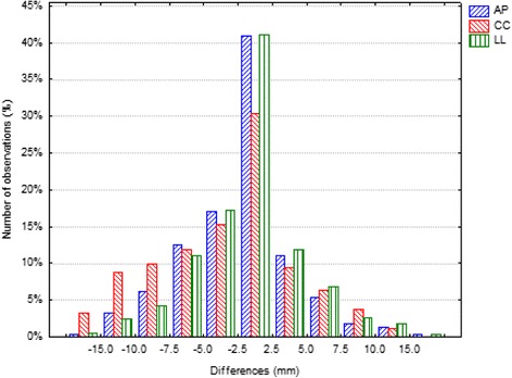
Forty patients with organ confined prostate cancer and candidates to curative radiotherapy were enrolled in this prospective study. At each treatment session, after laser alignment, all patients received imaging by a 3D-surface and a 3D-US system. The shifts along the three directions (anterior-posterior AP, cranial-caudal CC, and later-lateral LL) were measured in terms of systematic and random errors. Then, we performed statistical analysis on the differences and the possible correlations between the two modalities.
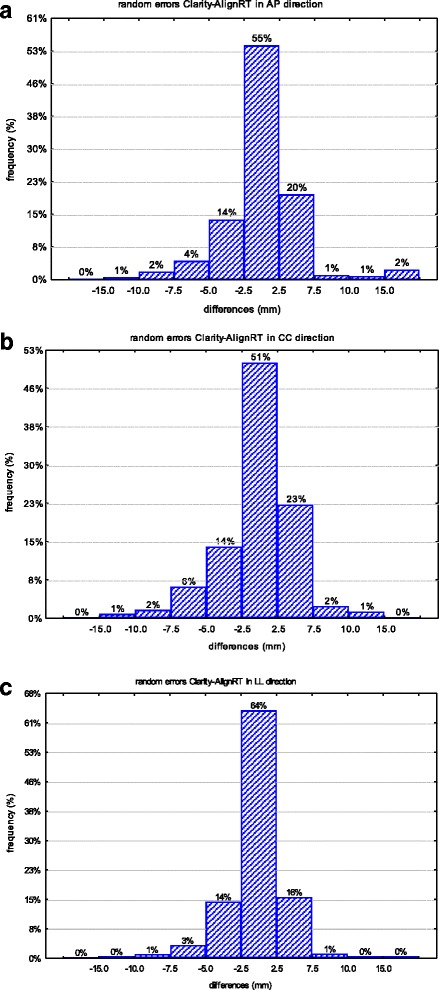
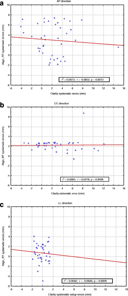
For both IGRT modalities, surface imaging and US, 1318 acquisitions were collected. According with Shapiro Wilk test, the positioning error distributions were not Gaussian for both modalities. The differences between the systematic errors detected by the two modalities were statistically significant only in LL direction (p < 0.05), while the differences between the random errors were not statistically significant in any directions. The 95% confidence interval of the residual errors obtained by subtracting the random errors detected with surface images to those detected with US was included in the range from -7 mm to 7 mm corresponding to the minimum PTV margin adopted in AP direction in our clinical routine.
From our data, it emerges that setup misalignments measured by surface imaging can be predictive of US displacements after the adjustment for systematic errors. Moreover, surface imaging can detect setup errors predictive of registration errors measured by US. This data suggest that the two IGRT modalities could be considered as complementary to each other and could represent a daily "low-cost" and non-invasive IGRT modality in prostate cancer patients.
图像引导放射治疗(IGRT)是实施高精度放射治疗的一项基本前提条件。我们比较了两种非电离成像方式(表面成像和经腹超声,US)检测到的每日变化,以验证前列腺癌患者的摆位以及内部器官的变化情况。
40例患有器官局限性前列腺癌且适合根治性放射治疗的患者纳入了这项前瞻性研究。在每次治疗过程中,激光定位后,所有患者均接受三维表面成像系统和三维超声系统的成像检查。沿着三个方向(前后方向AP、头脚方向CC和左右方向LL)的位移通过系统误差和随机误差进行测量。然后,我们对两种成像方式之间的差异及可能存在的相关性进行了统计分析。
对于表面成像和超声这两种IGRT方式,共收集了1318次成像数据。根据 Shapiro Wilk 检验,两种成像方式的定位误差分布均不呈高斯分布。两种成像方式检测到的系统误差之间的差异仅在LL方向具有统计学意义(p < 0.05),而随机误差在任何方向上的差异均无统计学意义。用表面成像检测到的随机误差减去超声检测到的随机误差所得到的残余误差的95%置信区间包含在-7毫米至7毫米的范围内,这与我们临床常规中在AP方向采用的最小计划靶区(PTV)边界相对应。
从我们的数据可以看出,表面成像测量的摆位偏差在对系统误差进行校正后可预测超声检测到的位移。此外,表面成像能够检测出可预测超声测量的配准误差的摆位误差。这些数据表明,这两种IGRT方式可被视为相互补充,并且可能代表了前列腺癌患者每日“低成本”且无创的IGRT方式。