• 文献检索
  • 文档翻译
  • 深度研究
  • 学术资讯
  • Suppr Zotero 插件Zotero 插件
  • 邀请有礼
  • 套餐&价格
  • 历史记录
应用&插件
Suppr Zotero 插件Zotero 插件浏览器插件Mac 客户端Windows 客户端微信小程序
定价
高级版会员购买积分包购买API积分包
服务
文献检索文档翻译深度研究API 文档MCP 服务
关于我们
关于 Suppr公司介绍联系我们用户协议隐私条款
关注我们

Suppr 超能文献

核心技术专利:CN118964589B侵权必究
粤ICP备2023148730 号-1Suppr @ 2026

文献检索

告别复杂PubMed语法,用中文像聊天一样搜索,搜遍4000万医学文献。AI智能推荐,让科研检索更轻松。

立即免费搜索

文件翻译

保留排版,准确专业,支持PDF/Word/PPT等文件格式,支持 12+语言互译。

免费翻译文档

深度研究

AI帮你快速写综述,25分钟生成高质量综述,智能提取关键信息,辅助科研写作。

立即免费体验

标准剂量与低剂量CT方案在局限性肺病变评估中的应用:病变特征分析能力-iLEAD研究

Standard-dose vs. low-dose CT protocols in the evaluation of localized lung lesions: Capability for lesion characterization-iLEAD study.

作者信息

Kubo Takeshi, Ohno Yoshiharu, Takenaka Daisuke, Nishino Mizuki, Gautam Shiva, Sugimura Kazuro, Kauczor Hans Ulrich, Hatabu Hiroto

机构信息

Department of Diagnostic Imaging and Nuclear Medicine, Kyoto University Graduate School of Medicine, 54 Shogoin Kawahara-cho, Sakyo-ku, Kyoto 606-8507, Japan.

Department of Radiology, Kobe University Graduate School of Medicine, 7-5-2 Kusunoki-cho, Chuo-ku, Kobe 650-0017, Japan.

出版信息

Eur J Radiol Open. 2016 Mar 24;3:67-73. doi: 10.1016/j.ejro.2016.03.002. eCollection 2016.

DOI:10.1016/j.ejro.2016.03.002
PMID:27957516
原文链接:https://pmc.ncbi.nlm.nih.gov/articles/PMC5144111/
Abstract

OBJECTIVE

To determine the lesion characterization capability by low dose CT for localized lung lesions in comparison with standard dose CT.

SUBJECTS AND METHODS

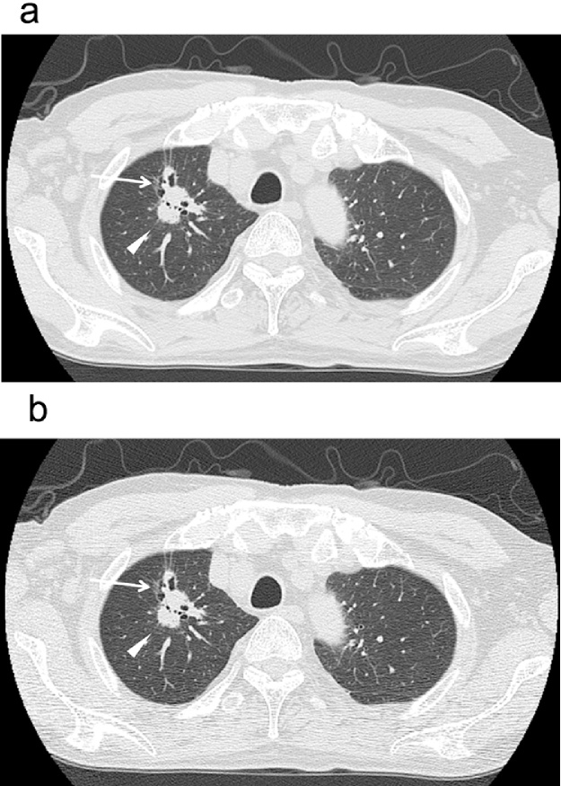
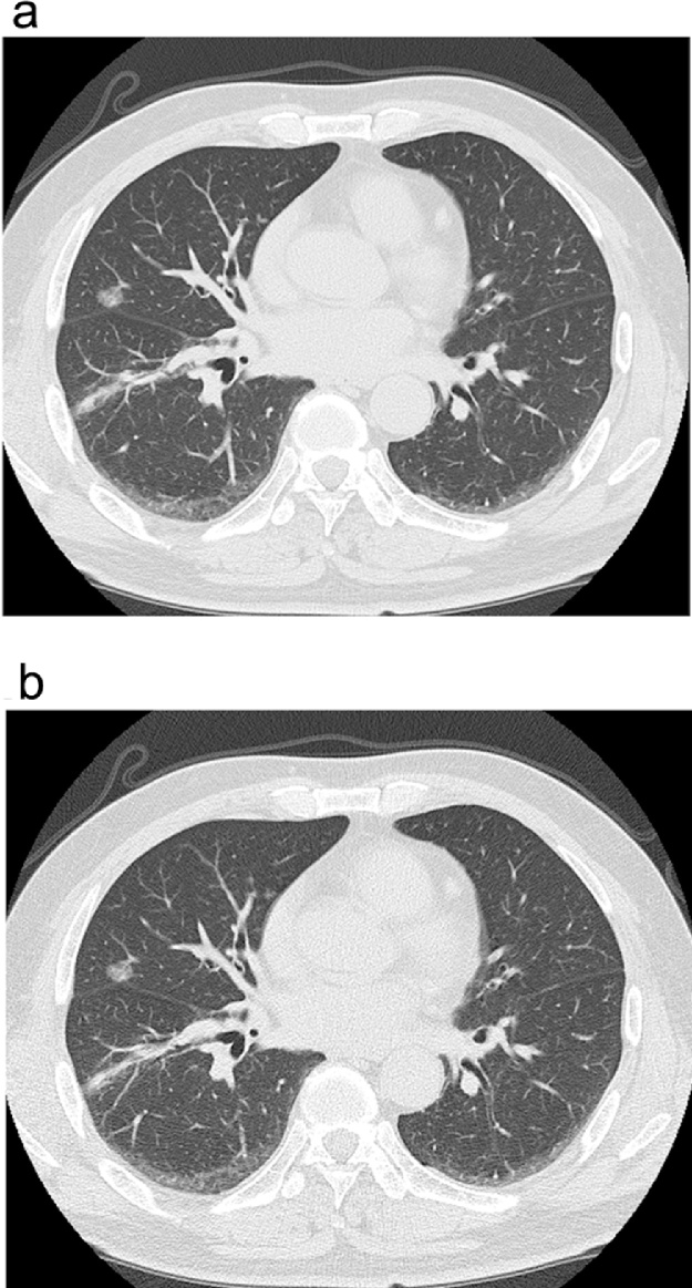
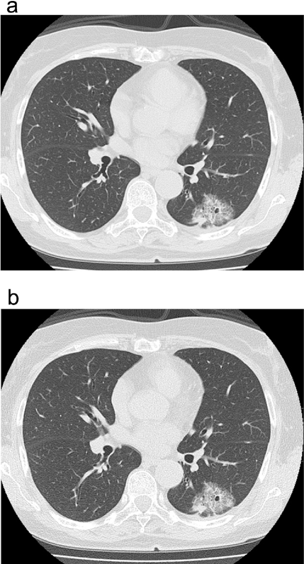
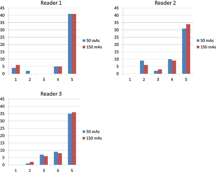
Approval for this study was granted by our Institutional Review Board. Fifty-two consecutive patients (36 males and 16 females, median age of 71 years.) who had CT examinations for evaluation of lung lesions comprise the study population. Two chest CT scans were performed with current time product of 50 and 150 mAs at 120 kVp, with the same scan length with a 16 detector-row CT scanner. Three readers evaluated 52 target lesions and assigned an overall impression score to each target lesion, using a 5 point scale from 1 (definitely benign) to 5 (definitely malignant). Six features of the lesions including lesion type, margin characteristics, calcification, lobulation, speculation, and pleural indentation were also reported with 5-point scales. The weighted kappa analyses and receiver operating characteristic analysis were used for analysis.

RESULTS

The mean kappa value between low-and standard-dose CT was 0.82 for overall impression of the lesions, showing almost perfect agreement. Area under the curve of low-dose CT (Az = 0.74) had no significant difference from that of standard-dose CT (Az = 0.74) (p = 0.61). The kappa values for six lesion features ranged from 0.45 to 0.83, showing moderate to almost perfect agreement.

CONCLUSION

Lesion characterization capability by low-dose CT images was comparable to that by standard-dose CT images and therefore sufficient for evaluation of localized lung lesions.

摘要

目的

与标准剂量CT相比,确定低剂量CT对局限性肺病变的病变特征描述能力。

对象与方法

本研究获得了我院机构审查委员会的批准。连续52例因评估肺病变而接受CT检查的患者(36例男性和16例女性,中位年龄71岁)组成了研究人群。使用16排探测器CT扫描仪,在120 kVp下分别以50和150 mAs的管电流时间乘积进行两次胸部CT扫描,扫描长度相同。三位阅片者对52个目标病变进行评估,并使用从1(肯定为良性)到5(肯定为恶性)的5分制为每个目标病变给出总体印象评分。还对病变的六个特征,包括病变类型、边缘特征、钙化、分叶、毛刺和胸膜凹陷,采用5分制进行报告。采用加权kappa分析和受试者操作特征分析进行分析。

结果

低剂量CT与标准剂量CT之间病变总体印象的平均kappa值为0.82,显示出几乎完全一致。低剂量CT的曲线下面积(Az = 0.74)与标准剂量CT的曲线下面积(Az = 0.74)无显著差异(p = 0.61)。六个病变特征的kappa值范围为0.45至0.83,显示出中度至几乎完全一致。

结论

低剂量CT图像的病变特征描述能力与标准剂量CT图像相当,因此足以用于评估局限性肺病变。

https://cdn.ncbi.nlm.nih.gov/pmc/blobs/dd95/5144111/977beaf2782e/gr6.jpg
https://cdn.ncbi.nlm.nih.gov/pmc/blobs/dd95/5144111/b1aaffc1ea65/gr1.jpg
https://cdn.ncbi.nlm.nih.gov/pmc/blobs/dd95/5144111/fe62e9b21dfe/gr2.jpg
https://cdn.ncbi.nlm.nih.gov/pmc/blobs/dd95/5144111/ea5ab50d11a9/gr3.jpg
https://cdn.ncbi.nlm.nih.gov/pmc/blobs/dd95/5144111/150c3b51ad56/gr4.jpg
https://cdn.ncbi.nlm.nih.gov/pmc/blobs/dd95/5144111/f007be48202f/gr5.jpg
https://cdn.ncbi.nlm.nih.gov/pmc/blobs/dd95/5144111/977beaf2782e/gr6.jpg

相似文献

1
Standard-dose vs. low-dose CT protocols in the evaluation of localized lung lesions: Capability for lesion characterization-iLEAD study.标准剂量与低剂量CT方案在局限性肺病变评估中的应用:病变特征分析能力-iLEAD研究
Eur J Radiol Open. 2016 Mar 24;3:67-73. doi: 10.1016/j.ejro.2016.03.002. eCollection 2016.
2
Low dose chest CT protocol (50 mAs) as a routine protocol for comprehensive assessment of intrathoracic abnormality.低剂量胸部CT方案(50毫安秒)作为全面评估胸内异常的常规方案。
Eur J Radiol Open. 2016 Apr 27;3:86-94. doi: 10.1016/j.ejro.2016.04.001. eCollection 2016.
3
Evaluation of thoracic abnormalities on 64-row multi-detector row CT: comparison between axial images versus coronal reformations.64排多层螺旋CT对胸部异常的评估:轴位图像与冠状位重组图像的比较
Eur J Radiol. 2006 Jul;59(1):33-41. doi: 10.1016/j.ejrad.2005.12.035. Epub 2006 Feb 15.
4
Quantitative Features of Liver Lesions, Lung Nodules, and Renal Stones at Multi-Detector Row CT Examinations: Dependency on Radiation Dose and Reconstruction Algorithm.多排螺旋 CT 检查中肝脏病变、肺结节和肾结石的定量特征:与辐射剂量和重建算法的关系。
Radiology. 2016 Apr;279(1):185-94. doi: 10.1148/radiol.2015150892. Epub 2015 Dec 1.
5
Combination of free-breathing radial 3D fat-suppressed T1-weighted gradient-echo sequence with diffusion weighted images: Potential for differentiating malignant from benign peripheral solid pulmonary masses.自由呼吸径向三维脂肪抑制T1加权梯度回波序列与扩散加权成像相结合:鉴别周围型实性肺肿块良恶性的潜力
Magn Reson Imaging. 2019 Apr;57:271-276. doi: 10.1016/j.mri.2018.12.004. Epub 2018 Dec 14.
6
High-pitch, 120 kVp/30 mAs, low-dose dual-source chest CT with iterative reconstruction: Prospective evaluation of radiation dose reduction and image quality compared with those of standard-pitch low-dose chest CT in healthy adult volunteers.高千伏、120kVp/30mAs、低剂量双源胸部 CT 迭代重建:前瞻性评估与标准管电压低剂量胸部 CT 比较在健康成年志愿者中的辐射剂量降低和图像质量。
PLoS One. 2019 Jan 24;14(1):e0211097. doi: 10.1371/journal.pone.0211097. eCollection 2019.
7
Standard-, Reduced-, and No-Dose Thin-Section Radiologic Examinations: Comparison of Capability for Nodule Detection and Nodule Type Assessment in Patients Suspected of Having Pulmonary Nodules.标准剂量、低剂量和零剂量薄层放射学检查:在疑似肺部结节患者中对结节检测和结节类型评估能力的比较。
Radiology. 2017 Aug;284(2):562-573. doi: 10.1148/radiol.2017161037. Epub 2017 Mar 6.
8
Suspected acute colon diverticulitis: imaging with low-dose unenhanced multi-detector row CT.疑似急性结肠憩室炎:低剂量非增强多层螺旋CT成像
Radiology. 2005 Oct;237(1):189-96. doi: 10.1148/radiol.2371041432. Epub 2005 Aug 26.
9
Pulmonary high-resolution ultrashort TE MR imaging: Comparison with thin-section standard- and low-dose computed tomography for the assessment of pulmonary parenchyma diseases.肺部高分辨率超短回波时间磁共振成像:与薄层标准剂量和低剂量计算机断层扫描在评估肺实质疾病中的比较。
J Magn Reson Imaging. 2016 Feb;43(2):512-32. doi: 10.1002/jmri.25008. Epub 2015 Jul 30.
10
Lung nodule detection performance in five observers on computed tomography (CT) with adaptive iterative dose reduction using three-dimensional processing (AIDR 3D) in a Japanese multicenter study: Comparison between ultra-low-dose CT and low-dose CT by receiver-operating characteristic analysis.日本多中心研究中五名观察者在使用三维处理的自适应迭代剂量降低技术(AIDR 3D)的计算机断层扫描(CT)上对肺结节的检测性能:通过接受者操作特征分析比较超低剂量CT和低剂量CT。
Eur J Radiol. 2015 Jul;84(7):1401-12. doi: 10.1016/j.ejrad.2015.03.012. Epub 2015 Apr 2.

引用本文的文献

1
Impact of a deep-learning image reconstruction algorithm on image quality and detection of solid lung lesions.深度学习图像重建算法对肺部实性病变图像质量及检测的影响
Res Diagn Interv Imaging. 2025 May 27;14:100062. doi: 10.1016/j.redii.2025.100062. eCollection 2025 Jun.
2
Low Dose Contrast Enhanced CT Thorax Protocol: Comparison of Low Kilovoltage, Low Contrast Volume Using Iterative Reconstruction Technique with Standard Protocol.低剂量对比增强胸部CT方案:采用迭代重建技术的低千伏、低对比剂用量与标准方案的比较
Ethiop J Health Sci. 2025 Mar;35(2):74-82. doi: 10.4314/ejhs.v35i2.2.
3
Deep Learning-Based Reconstruction Algorithm With Lung Enhancement Filter for Chest CT: Effect on Image Quality and Ground Glass Nodule Sharpness.

本文引用的文献

1
Radiation dose reduction in chest CT--review of available options.胸部CT辐射剂量的降低——现有方法综述
Eur J Radiol. 2014 Oct;83(10):1953-61. doi: 10.1016/j.ejrad.2014.06.033. Epub 2014 Jul 9.
2
Results of initial low-dose computed tomographic screening for lung cancer.肺癌初始低剂量计算机断层扫描筛查结果。
N Engl J Med. 2013 May 23;368(21):1980-91. doi: 10.1056/NEJMoa1209120.
3
Lung cancer screening, radiation, risks, benefits, and uncertainty.肺癌筛查、辐射、风险、益处及不确定性。
基于深度学习的带肺增强滤波器的胸部 CT 重建算法:对图像质量和磨玻璃结节锐利度的影响。
Korean J Radiol. 2024 Sep;25(9):833-842. doi: 10.3348/kjr.2024.0472.
4
Accuracy of patient-specific CT organ doses from Monte Carlo simulations: influence of CT-based voxel models.基于 CT 体素模型的蒙特卡罗模拟计算患者特定 CT 器官剂量的准确性研究。
Phys Eng Sci Med. 2024 Sep;47(3):989-1000. doi: 10.1007/s13246-024-01422-z. Epub 2024 Apr 18.
5
Deep Learning Image Reconstruction for CT: Technical Principles and Clinical Prospects.深度学习在 CT 图像重建中的应用:技术原理与临床前景。
Radiology. 2023 Mar;306(3):e221257. doi: 10.1148/radiol.221257. Epub 2023 Jan 31.
6
Sequential low-dose CT thorax scans to determine invasive pulmonary fungal infection incidence after allogeneic hematopoietic cell transplantation.序贯低剂量 CT 胸部扫描在异基因造血细胞移植后用于确定侵袭性肺部真菌感染的发生率。
Ann Hematol. 2023 Feb;102(2):413-420. doi: 10.1007/s00277-022-05062-9. Epub 2022 Dec 3.
7
Image Recovery from Synthetic Noise Artifacts in CT Scans Using Modified U-Net.基于改进 U-Net 的 CT 扫描合成噪声伪影图像恢复。
Sensors (Basel). 2022 Sep 16;22(18):7031. doi: 10.3390/s22187031.
8
A Task-dependent Investigation on Dose and Texture in CT Image Reconstruction.CT图像重建中剂量与纹理的任务相关研究。
IEEE Trans Radiat Plasma Med Sci. 2020 Jul;4(4):441-449. doi: 10.1109/trpms.2019.2957459. Epub 2019 Dec 4.
9
Baseline Chest Computed Tomography as Standard of Care in High-Risk Hematology Patients.基线胸部计算机断层扫描作为高危血液学患者的标准治疗手段
J Fungi (Basel). 2020 Mar 13;6(1):36. doi: 10.3390/jof6010036.
10
Video-assisted thoracoscopic surgery for invasive pulmonary fungal infection in haematology patients.血液科患者侵袭性肺真菌感染的电视辅助胸腔镜手术
J Thorac Dis. 2019 Jul;11(7):2839-2845. doi: 10.21037/jtd.2019.07.13.
JAMA. 2012 Jun 13;307(22):2434-5. doi: 10.1001/jama.2012.6096.
4
Reduced lung-cancer mortality with low-dose computed tomographic screening.低剂量计算机断层扫描筛查可降低肺癌死亡率。
N Engl J Med. 2011 Aug 4;365(5):395-409. doi: 10.1056/NEJMoa1102873. Epub 2011 Jun 29.
5
Detection of nodules showing ground-glass opacity in the lungs at low-dose multidetector computed tomography: phantom and clinical study.低剂量多层螺旋计算机断层扫描检测肺部磨玻璃样结节:体模与临床研究
J Comput Assist Tomogr. 2009 Jan-Feb;33(1):49-53. doi: 10.1097/RCT.0b013e31815e6291.
6
Use of 3D adaptive raw-data filter in CT of the lung: effect on radiation dose reduction.
AJR Am J Roentgenol. 2008 Oct;191(4):1071. doi: 10.2214/AJR/07.2630.
7
Radiation dose reduction in chest CT: a review.胸部CT辐射剂量降低:综述
AJR Am J Roentgenol. 2008 Feb;190(2):335-43. doi: 10.2214/AJR.07.2556.
8
Computed tomography--an increasing source of radiation exposure.计算机断层扫描——辐射暴露的一个日益增加的来源。
N Engl J Med. 2007 Nov 29;357(22):2277-84. doi: 10.1056/NEJMra072149.
9
3-dimensional adaptive raw-data filter: evaluation in low dose chest multidetector-row computed tomography.
J Comput Assist Tomogr. 2006 Nov-Dec;30(6):933-8. doi: 10.1097/01.rct.0000221951.60393.64.
10
Chest CT performed with z-axis modulation: scanning protocol and radiation dose.采用z轴调制进行的胸部CT:扫描方案与辐射剂量
Radiology. 2005 Oct;237(1):303-8. doi: 10.1148/radiol.2371041227.