Canessa Andrea, Pozzi Nicolò G, Arnulfo Gabriele, Brumberg Joachim, Reich Martin M, Pezzoli Gianni, Ghilardi Maria F, Matthies Cordula, Steigerwald Frank, Volkmann Jens, Isaias Ioannis U
Department of Neurology, University Hospital and Julius-Maximilian-University Wuerzburg, Germany.
Department of Nuclear Medicine, University Hospital and Julius-Maximilian-University Wuerzburg, Germany.
Front Hum Neurosci. 2016 Dec 6;10:611. doi: 10.3389/fnhum.2016.00611. eCollection 2016.
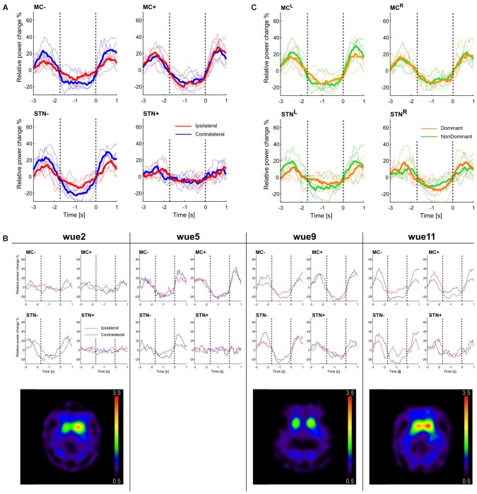
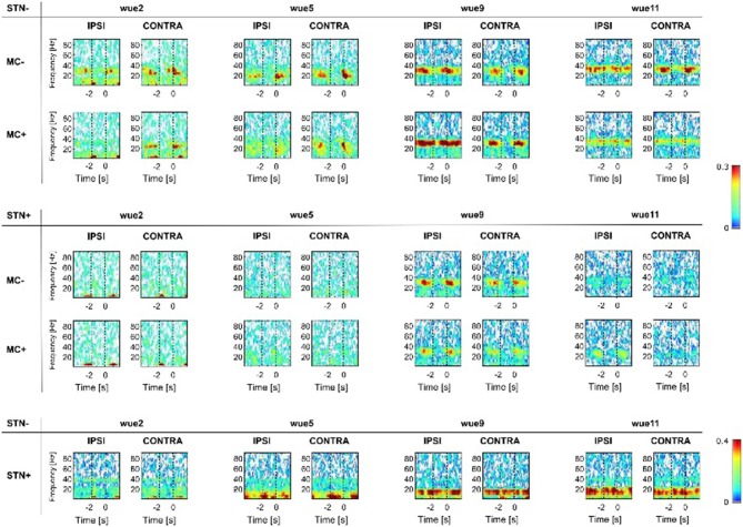
Activation of the basal ganglia has been shown during the preparation and execution of movement. However, the functional interaction of cortical and subcortical brain areas during movement and the relative contribution of dopaminergic striatal innervation remains unclear. We recorded local field potential (LFP) activity from the subthalamic nucleus (STN) and high-density electroencephalography (EEG) signals in four patients with Parkinson's disease (PD) off dopaminergic medication during a multi-joint motor task performed with their dominant and non-dominant hand. Recordings were performed by means of a fully-implantable deep brain stimulation (DBS) device at 4 months after surgery. Three patients also performed a single-photon computed tomography (SPECT) with [I]N-ω-fluoropropyl-2β-carbomethoxy-3β-(4-iodophenyl)nortropane (FP-CIT) to assess striatal dopaminergic innervation. Unilateral movement execution led to event-related desynchronization (ERD) followed by a rebound after movement termination event-related synchronization (ERS) of oscillatory beta activity in the STN and primary sensorimotor cortex of both hemispheres. Dopamine deficiency directly influenced movement-related beta-modulation, with greater beta-suppression in the most dopamine-depleted hemisphere for both ipsi- and contralateral hand movements. Cortical-subcortical, but not interhemispheric subcortical coherencies were modulated by movement and influenced by striatal dopaminergic innervation, being stronger in the most dopamine-depleted hemisphere. The data are consistent with a role of dopamine in shielding subcortical structures from an excessive cortical entrapment and cross-hemispheric coupling, thus allowing fine-tuning of movement.
在运动准备和执行过程中,基底神经节已被证明处于激活状态。然而,运动过程中皮质和皮质下脑区的功能相互作用以及多巴胺能纹状体神经支配的相对贡献仍不清楚。我们记录了4例未服用多巴胺能药物的帕金森病(PD)患者在使用优势手和非优势手进行多关节运动任务期间丘脑底核(STN)的局部场电位(LFP)活动和高密度脑电图(EEG)信号。记录通过完全植入式深部脑刺激(DBS)装置在术后4个月进行。3例患者还进行了单光子计算机断层扫描(SPECT),使用[I]N-ω-氟丙基-2β-甲氧基羰基-3β-(4-碘苯基)去甲托烷(FP-CIT)来评估纹状体多巴胺能神经支配。单侧运动执行导致事件相关去同步化(ERD),随后在运动终止后出现振荡β活动的反弹,即事件相关同步化(ERS),在两个半球的STN和初级感觉运动皮层均有出现。多巴胺缺乏直接影响与运动相关的β调制,在多巴胺耗竭最严重的半球,同侧和对侧手部运动的β抑制作用更强。皮质-皮质下相干性而非半球间皮质下相干性受运动调制,并受纹状体多巴胺能神经支配影响,在多巴胺耗竭最严重的半球更强。这些数据与多巴胺在保护皮质下结构免受过度皮质束缚和跨半球耦合方面的作用一致,从而允许对运动进行微调。