Salvatore Viviana, Teti Gabriella, Focaroli Stefano, Mazzotti Maria Carla, Mazzotti Antonio, Falconi Mirella
DIBINEM, University of Bologna Department of Biomedical and Neuromotor Sciences, 40126 Bologna, Italy.
DIMEC, University of Bologna, Department of Medical and Surgical Sciences, 40126 Bologna, Italy.
Oncotarget. 2017 Feb 7;8(6):9608-9616. doi: 10.18632/oncotarget.14155.
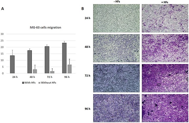
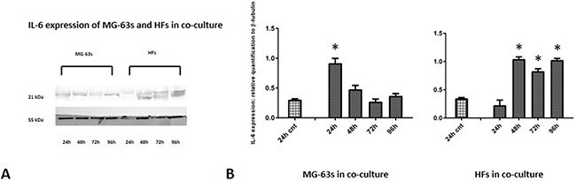
The tumor microenvironment contributes to cancer progression, in part through interactions between tumor and normal stromal cells. This study analyzed morphological and molecular changes induced in co-cultured human fibroblasts (HFs) and the MG-63 osteosarcoma cell line. Co-cultured cell monolayers were morphologically analyzed using high resolution scanning electron microscopy (HR-SEM), and trans-well assays were performed to assess cell migration and invasion. Proteins involved in inflammatory responses, cancer cell invasion, and angiogenesis were assessed using western blotting. HR-SEM showed progressive spatial orientation loss by fibroblasts in contact with MG-63s, while MG-63s proliferated rapidly and invaded HF space. Trans-well assays showed enhanced MG-63 migration in the presence of HFs. IL-6 expression was increased in co-cultured HFs, possibly stimulated by TNF-α. HFs do not normally express YKL-40 but did so in co-culture. Band densitometric analyses showed that increasing YKL-40 expression was followed by VEGF overexpression, especially in MG-63s. Finally, our results confirmed fibroblasts as the main matrix metalloproteinase source in this tumor microenvironment. Our study sheds new light on how tumor-stroma interactions promote tumor development and progression, and may support identification of novel anti-cancer therapeutics.
肿瘤微环境在一定程度上通过肿瘤细胞与正常基质细胞之间的相互作用促进癌症进展。本研究分析了共培养的人成纤维细胞(HFs)和MG-63骨肉瘤细胞系中诱导的形态学和分子变化。使用高分辨率扫描电子显微镜(HR-SEM)对共培养的细胞单层进行形态学分析,并进行Trans-well实验以评估细胞迁移和侵袭能力。通过蛋白质印迹法评估参与炎症反应、癌细胞侵袭和血管生成的蛋白质。HR-SEM显示,与MG-63细胞接触的成纤维细胞逐渐失去空间定向,而MG-63细胞迅速增殖并侵入HFs空间。Trans-well实验表明,在有HFs存在的情况下,MG-63细胞的迁移能力增强。共培养的HFs中IL-6表达增加,可能是由TNF-α刺激所致。HFs通常不表达YKL-40,但在共培养时表达。条带密度分析显示,YKL-40表达增加后VEGF过表达,尤其是在MG-63细胞中。最后,我们的结果证实成纤维细胞是这种肿瘤微环境中主要的基质金属蛋白酶来源。我们的研究为肿瘤-基质相互作用如何促进肿瘤发展和进展提供了新的线索,并可能有助于确定新的抗癌治疗方法。