Siddiqui Zeba Rahman, Jhingran Rajesh, Bains Vivek Kumar, Srivastava Ruchi, Madan Rohit, Rizvi Iram
Department of Periodontology, Saraswati Dental College and Hospital, Lucknow, Uttar Pradesh, India.
Eur J Dent. 2016 Oct-Dec;10(4):496-506. doi: 10.4103/1305-7456.195160.
The objective of the study was to evaluate clinically and radiographically the efficacy of platelet-rich fibrin (PRF) versus β-tri-calcium phosphate (β-TCP) in the treatment of Grade II mandibular furcation defects.
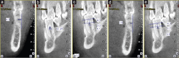
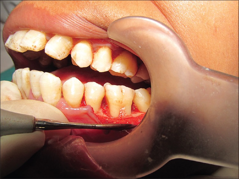
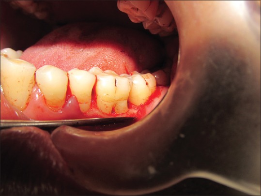
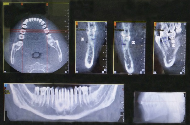
Forty-five Grade II furcation defect in mandibular molars which were assigned to open flap debridement (OFD) with PRF Group I ( = 15), to OFD with β-TCP Group II ( = 15), and to OFD alone Group III ( = 15) were analyzed for clinical parameters (probing pocket depth [PPD], vertical clinical attachment level [VCAL], horizontal clinical attachment level [HCAL], gingival recession, relative vertical height of furcation [r-VHF], and relative horizontal depth of furcation [r-HDF]) and radiographical parameters (horizontal depth of furcation [H-DOF], vertical height of furcation [V-HOF]) using cone-beam computed tomography (CBCT) at 6 months interval.
For clinical parameters, reduction in PPD and gain in VCAL and HCAL were higher in Group II as compared to Group I. Change in r-VHF and r-HDF was greater in Group II as compared to Group I. Mean percentage clinical vertical defect fill was higher in Group II as compared to Group I (58.52% ± 11.68% vs. 53.24% ± 13.22%, respectively). On CBCT, mean change at 6 months for all parameters showed nonsignificant difference between the two experimental groups. Mean change in V-HOF was higher in Group I as compared to Group II, but mean change in H-DOF and furcation width was more in Group II as compared to Group I.
For both experimental and control groups, there was statistically significant improvement at 6 months follow-up from baseline values.
本研究旨在从临床和影像学方面评估富血小板纤维蛋白(PRF)与β-磷酸三钙(β-TCP)治疗下颌Ⅱ度根分叉病变的疗效。
将45例下颌磨牙Ⅱ度根分叉病变患者分为三组,每组15例。第一组采用开放瓣清创术(OFD)联合PRF治疗,第二组采用OFD联合β-TCP治疗,第三组仅采用OFD治疗。分别于术后6个月时,采用锥形束计算机断层扫描(CBCT)分析临床参数(探诊深度[PPD]、垂直临床附着水平[VCAL]、水平临床附着水平[HCAL]、牙龈退缩、根分叉相对垂直高度[r-VHF]和根分叉相对水平深度[r-HDF])和影像学参数(根分叉水平深度[H-DOF]、根分叉垂直高度[V-HOF])。
临床参数方面,与第一组相比,第二组PPD降低、VCAL和HCAL增加更为明显。与第一组相比,第二组r-VHF和r-HDF的变化更大。第二组平均临床垂直缺损填充百分比高于第一组(分别为58.52%±11.68%和53.24%±13.22%)。CBCT检查显示,术后6个月时两组所有参数的平均变化无显著差异。与第二组相比,第一组V-HOF平均变化更大,但与第一组相比,第二组H-DOF和根分叉宽度平均变化更大。
对于实验组和对照组,术后6个月随访时与基线值相比均有统计学意义的改善。