• 文献检索
  • 文档翻译
  • 深度研究
  • 学术资讯
  • Suppr Zotero 插件Zotero 插件
  • 邀请有礼
  • 套餐&价格
  • 历史记录
应用&插件
Suppr Zotero 插件Zotero 插件浏览器插件Mac 客户端Windows 客户端微信小程序
定价
高级版会员购买积分包购买API积分包
服务
文献检索文档翻译深度研究API 文档MCP 服务
关于我们
关于 Suppr公司介绍联系我们用户协议隐私条款
关注我们

Suppr 超能文献

核心技术专利:CN118964589B侵权必究
粤ICP备2023148730 号-1Suppr @ 2026

文献检索

告别复杂PubMed语法,用中文像聊天一样搜索,搜遍4000万医学文献。AI智能推荐,让科研检索更轻松。

立即免费搜索

文件翻译

保留排版,准确专业,支持PDF/Word/PPT等文件格式,支持 12+语言互译。

免费翻译文档

深度研究

AI帮你快速写综述,25分钟生成高质量综述,智能提取关键信息,辅助科研写作。

立即免费体验

研究并确定眼前节光学相干断层扫描和超声生物显微镜在角膜及结膜肿瘤中的作用。

To Study and Determine the Role of Anterior Segment Optical Coherence Tomography and Ultrasound Biomicroscopy in Corneal and Conjunctival Tumors.

作者信息

Janssens Katleen, Mertens Michelle, Lauwers Noémie, de Keizer Rob J W, Mathysen Danny G P, De Groot Veva

机构信息

University of Antwerp, Antwerp, Belgium.

Department of Ophthalmology, University Hospital Antwerp, Edegem, Belgium.

出版信息

J Ophthalmol. 2016;2016:1048760. doi: 10.1155/2016/1048760. Epub 2016 Dec 6.

DOI:10.1155/2016/1048760
PMID:28050274
原文链接:https://pmc.ncbi.nlm.nih.gov/articles/PMC5168478/
Abstract

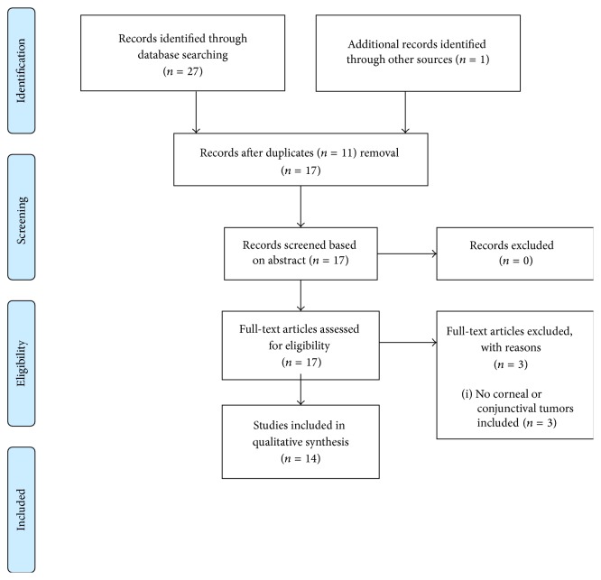
. To analyze and describe corneal and conjunctival tumor thickness and internal characteristics and extension in depth and size and shape measured by two noninvasive techniques, anterior segment optical coherence tomography (AS-OCT) and ultrasound biomicroscopy (UBM). . Systematic review. . This systematic review is based on a comprehensive search of 4 databases (Medline, Embase, Web of Science, and Cochrane Library). Articles published between January 1, 1999, and December 31, 2015, were included. We searched for articles using the following search terms in various combinations: "optical coherence tomography", "ultrasound biomicroscopy", "corneal neoplasm", "conjunctival neoplasm", "eye", "tumor" and "anterior segment tumors". Inclusion criteria were as follows: UBM and/or AS-OCT was used; the study included corneal or conjunctival tumors; and the article was published in English, French, Dutch, or German. . There were 14 sources selected. . Several studies on the quality of AS-OCT and UBM show that these imaging techniques provide useful information about the internal features, extension, size, and shape of tumors. Yet there is no enough evidence on the advantages and disadvantages of UBM and AS-OCT in certain tumor types. . More comparative studies are needed to investigate which imaging technique is most suitable for a certain tumor type.

摘要

通过两种非侵入性技术,即眼前节光学相干断层扫描(AS-OCT)和超声生物显微镜检查(UBM),分析并描述角膜和结膜肿瘤的厚度、内部特征以及在深度、大小和形状方面的扩展情况。

系统评价。

本系统评价基于对4个数据库(医学文献数据库、荷兰医学文摘数据库、科学引文索引数据库和考克兰图书馆)的全面检索。纳入了1999年1月1日至2015年12月31日期间发表的文章。我们使用以下检索词的各种组合来检索文章:“光学相干断层扫描”“超声生物显微镜检查”“角膜肿瘤”“结膜肿瘤”“眼”“肿瘤”和“眼前节肿瘤”。纳入标准如下:使用了UBM和/或AS-OCT;研究包括角膜或结膜肿瘤;文章以英文、法文、荷兰文或德文发表。

共选取了14篇文献。

几项关于AS-OCT和UBM质量的研究表明,这些成像技术可提供有关肿瘤内部特征、扩展、大小和形状的有用信息。然而,对于UBM和AS-OCT在某些肿瘤类型中的优缺点,尚无足够证据。

需要更多的比较研究来调查哪种成像技术最适合某一特定肿瘤类型。

https://cdn.ncbi.nlm.nih.gov/pmc/blobs/bf0e/5168478/c793a0012dd6/JOPH2016-1048760.001.jpg

相似文献

1
To Study and Determine the Role of Anterior Segment Optical Coherence Tomography and Ultrasound Biomicroscopy in Corneal and Conjunctival Tumors.研究并确定眼前节光学相干断层扫描和超声生物显微镜在角膜及结膜肿瘤中的作用。
J Ophthalmol. 2016;2016:1048760. doi: 10.1155/2016/1048760. Epub 2016 Dec 6.
2
Anterior segment optical coherence tomography and ultrasound biomicroscopy for measuring thickness of corneal and bulbar conjunctival tumours.眼前节光学相干断层扫描和超声生物显微镜测量角膜和球结膜肿瘤的厚度。
Br J Ophthalmol. 2022 Jun;106(6):760-764. doi: 10.1136/bjophthalmol-2018-312337. Epub 2021 Feb 5.
3
Comparison of high-resolution Scheimpflug and high-frequency ultrasound biomicroscopy to anterior-segment OCT corneal thickness measurements.高分辨率Scheimpflug成像与高频超声生物显微镜在前节光学相干断层扫描角膜厚度测量中的比较。
Clin Ophthalmol. 2013;7:2239-47. doi: 10.2147/OPTH.S53718. Epub 2013 Nov 20.
4
Correlation of anterior segment optical coherence tomography and ultrasound biomicroscopy in congenital corneal opacity.先天性角膜混浊的眼前节光学相干断层扫描与超声生物显微镜的相关性。
J AAPOS. 2024 Apr;28(2):103863. doi: 10.1016/j.jaapos.2024.103863. Epub 2024 Mar 6.
5
Comparison of anterior segment optical coherence tomography and ultrasound biomicroscopy for assessment of the anterior segment.用于眼前节评估的眼前节光学相干断层扫描与超声生物显微镜检查的比较
J Cataract Refract Surg. 2007 May;33(5):837-40. doi: 10.1016/j.jcrs.2007.01.021.
6
Conjunctival melanocytic naevus: Diagnostic value of anterior segment optical coherence tomography and ultrasound biomicroscopy.结膜黑素细胞痣:眼前节光学相干断层扫描和超声生物显微镜检查的诊断价值
PLoS One. 2018 Feb 14;13(2):e0192908. doi: 10.1371/journal.pone.0192908. eCollection 2018.
7
Anterior segment imaging for iris melanocytic tumors.虹膜黑素细胞肿瘤的眼前节成像
Eur J Ophthalmol. 2011 Sep-Oct;21(5):608-14. doi: 10.5301/EJO.2011.6214.
8
Spectral-domain optical coherence tomography versus ultrasound biomicroscopy for imaging of nonpigmented iris tumors.频域光相干断层扫描与超声生物显微镜在非色素性虹膜肿瘤成像中的比较。
Am J Ophthalmol. 2013 Oct;156(4):806-12. doi: 10.1016/j.ajo.2013.05.025. Epub 2013 Jul 20.
9
Optical coherence tomography and ultrasound biomicroscopy imaging of opaque corneas.光学相干断层扫描和超声生物显微镜成像在混浊角膜中的应用。
Cornea. 2013 Apr;32(4):e25-30. doi: 10.1097/ICO.0b013e318261eb2b.
10
Comparison of anterior chamber depth measurements by 3-dimensional optical coherence tomography, partial coherence interferometry biometry, Scheimpflug rotating camera imaging, and ultrasound biomicroscopy.三维光学相干断层扫描、部分相干干涉测量生物测量、Scheimpflug 旋转相机成像和超声生物显微镜测量前房深度的比较。
J Cataract Refract Surg. 2012 Jul;38(7):1207-13. doi: 10.1016/j.jcrs.2012.02.036. Epub 2012 May 19.

引用本文的文献

1
Preoperative Structural Risk Factors for Glaucoma After Penetrating Keratoplasty for Congenital Corneal Opacity: An Observational Study.先天性角膜混浊穿透性角膜移植术后青光眼的术前结构危险因素:一项观察性研究
Ophthalmol Ther. 2024 Nov;13(11):2869-2886. doi: 10.1007/s40123-024-01027-5. Epub 2024 Sep 13.
2
Anterior segment optical coherence tomography in ocular surface tumours and simulating lesions.眼前节光学相干断层扫描在眼表肿瘤和模拟病变中的应用。
Eye (Lond). 2023 Apr;37(5):925-937. doi: 10.1038/s41433-022-02339-1. Epub 2022 Dec 13.
3
Anterior segment optical coherence tomography angiography in the assessment of ocular surface lesions.

本文引用的文献

1
Anterior Segment Optical Coherence Tomography and its Clinical Applications in Glaucoma.眼前节光学相干断层扫描及其在青光眼的临床应用
J Curr Glaucoma Pract. 2012 May-Aug;6(2):68-74. doi: 10.5005/jp-journals-10008-1109. Epub 2012 Aug 16.
2
High-Resolution Optical Coherence Tomography as an Adjunctive Tool in the Diagnosis of Corneal and Conjunctival Pathology.高分辨率光学相干断层扫描作为角膜和结膜病变诊断的辅助工具。
Ocul Surf. 2015 Jul;13(3):226-35. doi: 10.1016/j.jtos.2015.02.001. Epub 2015 Apr 6.
3
[Comparison of thickness values in nine macular subfields using time-domain and spectral-domain optical coherence tomography].
眼前节光学相干断层扫描血管造影在眼表病变评估中的应用。
Ocul Surf. 2021 Oct;22:86-93. doi: 10.1016/j.jtos.2021.07.009. Epub 2021 Jul 30.
4
Deep Learning-Based Retinal Nerve Fiber Layer Thickness Measurement of Murine Eyes.基于深度学习的鼠眼视网膜神经纤维层厚度测量。
Transl Vis Sci Technol. 2021 Jul 1;10(8):21. doi: 10.1167/tvst.10.8.21.
5
Incidentally detected primary corneal squamous neoplasia in an elderly man presenting with cataract.偶然发现一位老年男性白内障患者存在原发性角膜鳞状上皮肿瘤。
BMJ Case Rep. 2020 Nov 9;13(11):e235789. doi: 10.1136/bcr-2020-235789.
6
The Effectiveness of Ultrasound Biomicroscopic and Anterior Segment Optical Coherence Tomography in the Assessment of Anterior Segment Tumors: Long-Term Follow-Up.超声生物显微镜和眼前节光学相干断层扫描在眼前节肿瘤评估中的有效性:长期随访
J Ophthalmol. 2020 Jun 16;2020:9053737. doi: 10.1155/2020/9053737. eCollection 2020.
7
AS-OCT Guided Treatment Of Diffuse Conjunctival Squamous Cell Carcinoma With Resection, Amniotic Membrane Graft And Topical Mitomycin C.光学相干断层扫描引导下采用切除、羊膜移植及局部丝裂霉素C治疗弥漫性结膜鳞状细胞癌
Clin Ophthalmol. 2019 Nov 20;13:2269-2278. doi: 10.2147/OPTH.S229399. eCollection 2019.
8
Assessment of Anterior Chamber by Ultrasound Biomicroscopy and Anterior Segment Optical Coherence Tomography in Patients with Inflammatory Glaucoma.超声生物显微镜和眼前节光学相干断层扫描对炎症性青光眼患者前房的评估
J Int Med Res. 2019 Dec;47(12):5950-5956. doi: 10.1177/0300060519867808. Epub 2019 Oct 8.
9
A novel use of ultrasound biomicroscopy.超声生物显微镜的一种新用途。
Eye (Lond). 2018 Feb;32(2):474-475. doi: 10.1038/eye.2017.190. Epub 2017 Sep 8.
[使用时域和频域光学相干断层扫描技术比较九个黄斑亚区的厚度值]
Orv Hetil. 2013 Dec 29;154(52):2059-64. doi: 10.1556/OH.2013.29785.
4
[Imaging of the iridocorneal angle in glaucoma].[青光眼患者前房角的影像学检查]
J Fr Ophtalmol. 2013 Apr;36(4):378-83. doi: 10.1016/j.jfo.2013.01.006. Epub 2013 Mar 29.
5
Diagnosis of ocular surface lesions using ultra-high-resolution optical coherence tomography.使用超高速光学相干断层扫描诊断眼表病变。
Ophthalmology. 2013 May;120(5):883-91. doi: 10.1016/j.ophtha.2012.10.025. Epub 2013 Jan 21.
6
The role of anterior segment optical coherence tomography in glaucoma.眼前节光学相干断层扫描在青光眼诊疗中的作用
J Ophthalmol. 2012;2012:476801. doi: 10.1155/2012/476801. Epub 2012 Aug 1.
7
Ultra-high resolution optical coherence tomography for differentiation of ocular surface squamous neoplasia and pterygia.用于区分眼表鳞状上皮肿瘤和翼状胬肉的超高分辨率光学相干断层扫描。
Ophthalmology. 2012 Mar;119(3):481-6. doi: 10.1016/j.ophtha.2011.08.028. Epub 2011 Dec 9.
8
Diagnosis and management of conjunctival and corneal intraepithelial neoplasia using ultra high-resolution optical coherence tomography.使用超高速分辨率光学相干断层扫描技术诊断和治疗结膜和角膜上皮内瘤变。
Ophthalmology. 2011 Aug;118(8):1531-7. doi: 10.1016/j.ophtha.2011.01.005. Epub 2011 Apr 20.
9
Pterygia measurements are more accurate with anterior segment optical coherence tomography - a pilot study.使用眼前节光学相干断层扫描测量翼状胬肉更准确——一项初步研究
Nepal J Ophthalmol. 2011 Jan-Jun;3(1):9-12. doi: 10.3126/nepjoph.v3i1.4271.
10
Assessment of anterior segment tumors with ultrasound biomicroscopy versus anterior segment optical coherence tomography in 200 cases.200 例前节肿瘤的超声生物显微镜与前节光学相干断层扫描评估。
Ophthalmology. 2011 Jul;118(7):1297-302. doi: 10.1016/j.ophtha.2010.11.011. Epub 2011 Mar 5.