Polans James, Cunefare David, Cole Eli, Keller Brenton, Mettu Priyatham S, Cousins Scott W, Allingham Michael J, Izatt Joseph A, Farsiu Sina
Opt Lett. 2017 Jan 1;42(1):17-20. doi: 10.1364/OL.42.000017.
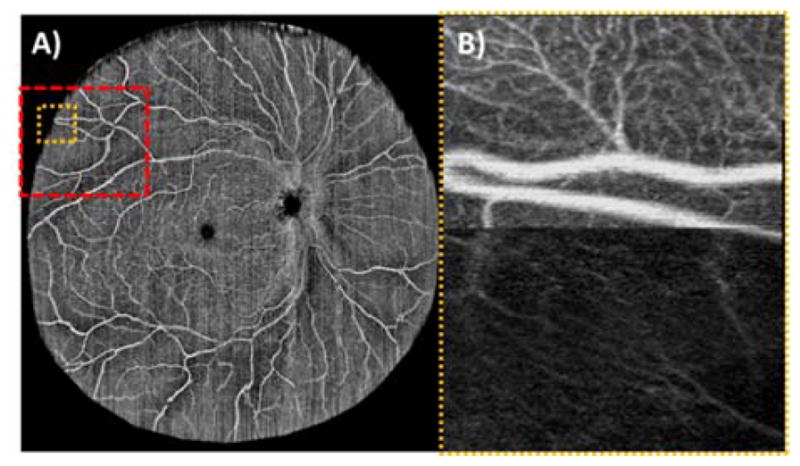
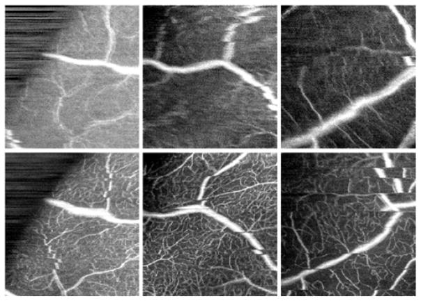
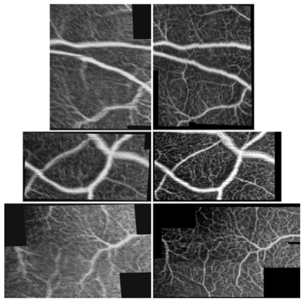
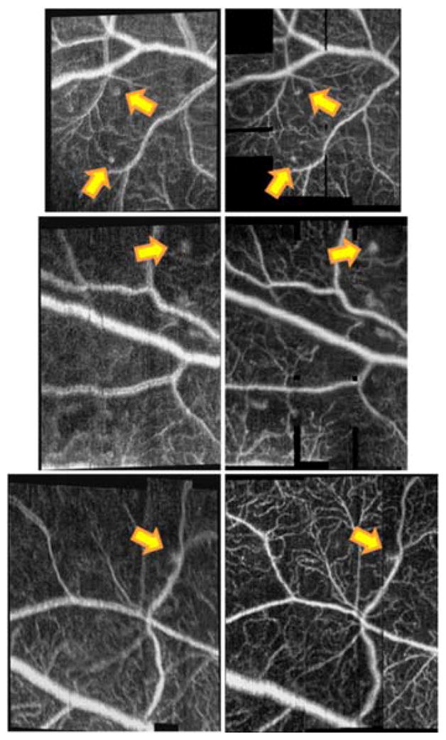
Optical coherence tomography angiography (OCTA) is a promising technique for non-invasive visualization of vessel networks in the human eye. We debut a system capable of acquiring wide field-of-view (>70°) OCT angiograms without mosaicking. Additionally, we report on enhancing the visualization of peripheral microvasculature using wavefront sensorless adaptive optics (WSAO). We employed a fast WSAO algorithm that enabled wavefront correction in <2 s by iterating the mirror shape at the speed of OCT B-scans rather than volumes. Also, we contrasted ∼7° field-of-view OCTA angiograms acquired in the periphery with and without WSAO correction. On average, WSAO improved the sharpness of microvasculature by 65% in healthy eyes and 38% in diseased eyes. Preliminary observations demonstrated that the location of 7° images could be identified directly from the wide field-of-view angiogram. A pilot study on a normal subject and patients with diabetic retinopathy showed the impact of utilizing WSAO for OCTA when visualizing peripheral vasculature pathologies.
光学相干断层扫描血管造影(OCTA)是一种用于无创可视化人眼血管网络的有前景的技术。我们首次推出了一种能够在不拼接的情况下获取宽视野(>70°)OCT血管造影的系统。此外,我们报告了使用无波前传感器自适应光学(WSAO)增强周边微血管系统的可视化。我们采用了一种快速WSAO算法,通过以OCT B扫描而非容积的速度迭代镜面形状,在<2秒内实现波前校正。此外,我们对比了在周边区域获取的有和没有WSAO校正的约7°视野OCTA血管造影。平均而言,WSAO使健康眼睛的微血管清晰度提高了65%,患病眼睛提高了38%。初步观察表明,可以直接从宽视野血管造影中识别7°图像的位置。一项针对正常受试者和糖尿病视网膜病变患者的初步研究显示了在可视化周边血管病变时利用WSAO进行OCTA的影响。