Kotlarek Katelyn J, Perry Jamie L, Fang Xiangming
East Carolina University, Greenville, NC.
J Craniofac Surg. 2017 May;28(3):833-837. doi: 10.1097/SCS.0000000000003373.
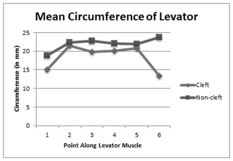
The purpose of this study was to examine differences in levator veli palatini (levator) morphology between adults with repaired cleft palate and adults with noncleft anatomy. Fifteen adult participants (10 with noncleft anatomy, 5 with repaired cleft palate) underwent 3-dimensional (3D) static magnetic resonance imaging (MRI). Image analyses included measures of total muscle volume and the circumference and diameter at 6 points along the length of the muscle. Differences between groups were analyzed using independent sample Mann-Whitney U tests (α < 0.05). Significant differences between groups were noted for measures of muscle volume, circumference at the origin and midline, anterior-posterior diameter at the origin and midline, and superior-inferior diameter at the point of insertion into the velum and midline. Differences in measures at other points along the levator muscle belly were not statistically significant. Limited sample size and gender differences may have impacted statistical findings. Overall, the levator muscle in adults with repaired cleft palate is significantly different than that of adults with noncleft anatomy. This study demonstrates the successful implementation of a method for 3D analysis of velopharyngeal (VP) musculature with potential clinical utility given continued technological advancements in MRI. Continued evaluation of pre- and postsurgical anatomy and short- and long-term outcomes may contribute to a better understanding of the effects of various types of palatoplasties on levator structure, which is important to VP function for speech.
本研究的目的是检查腭裂修复术后成人与非腭裂成人之间腭帆提肌形态的差异。15名成年参与者(10名非腭裂者,5名腭裂修复者)接受了三维(3D)静态磁共振成像(MRI)检查。图像分析包括测量肌肉总体积以及沿肌肉长度6个点的周长和直径。使用独立样本曼-惠特尼U检验(α<0.05)分析组间差异。在肌肉体积、起点和中线处的周长、起点和中线处的前后径以及插入软腭和中线处的上下径测量方面,组间存在显著差异。沿腭帆提肌肌腹其他点的测量差异无统计学意义。样本量有限和性别差异可能影响了统计结果。总体而言,腭裂修复术后成人的腭帆提肌与非腭裂成人的腭帆提肌有显著差异。鉴于MRI技术的不断进步,本研究证明了一种用于腭咽(VP)肌肉组织三维分析方法的成功实施及其潜在的临床应用价值。对手术前后解剖结构以及短期和长期结果的持续评估可能有助于更好地理解各种腭裂修复术对腭帆提肌结构的影响,这对语音的VP功能很重要。