Yan Dongmei, Zhang Zhihong, Luo Qingming, Yang Xiaoquan
Britton Chance Center for Biomedical Photonics, Wuhan National Laboratory for Optoelectronics-Huazhong University of Science and Technology, Wuhan, Hubei, China.
Key Laboratory of Biomedical Photonics of Ministry of Education, Huazhong University of Science and Technology, Wuhan, Hubei, China.
PLoS One. 2017 Jan 6;12(1):e0169424. doi: 10.1371/journal.pone.0169424. eCollection 2017.
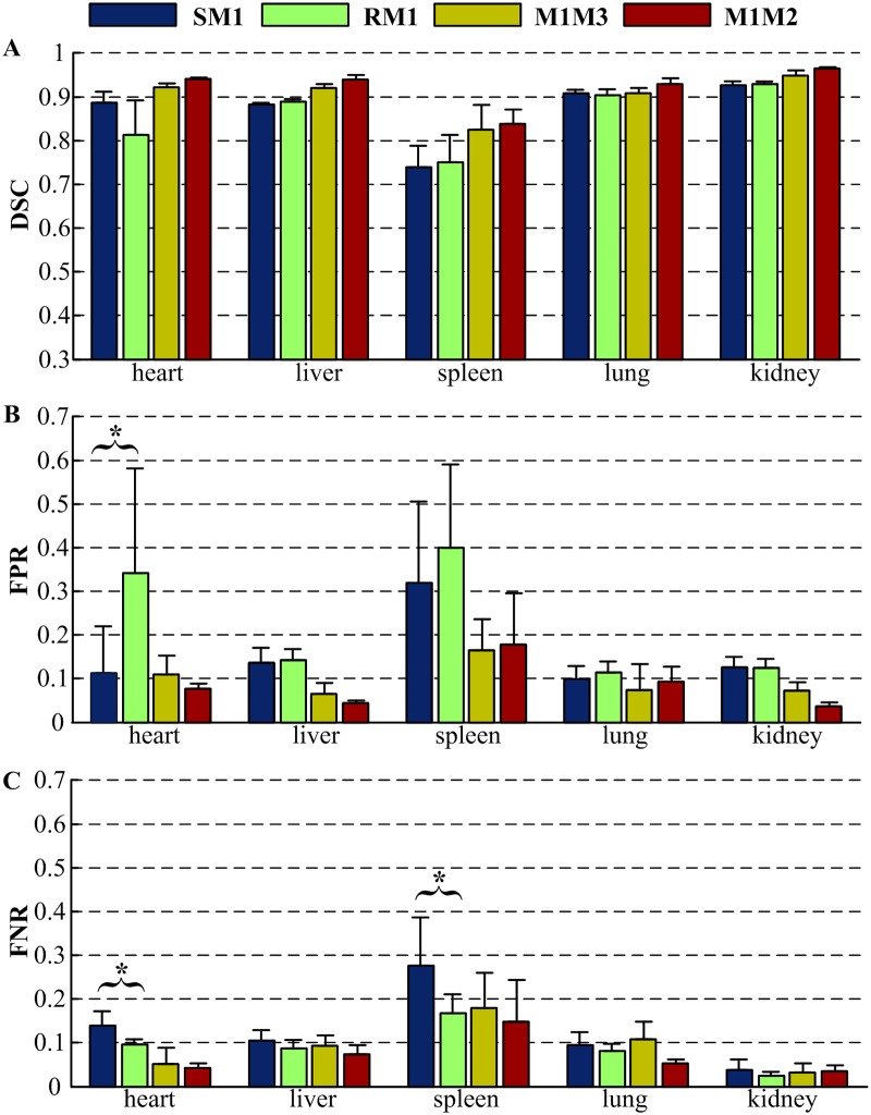
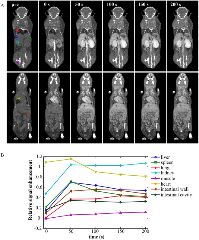
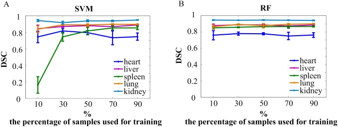
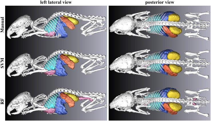
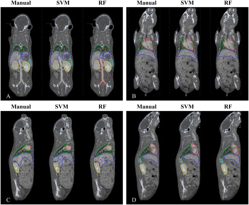
With the development of hybrid imaging scanners, micro-CT is widely used in locating abnormalities, studying drug metabolism, and providing structural priors to aid image reconstruction in functional imaging. Due to the low contrast of soft tissues, segmentation of soft tissue organs from mouse micro-CT images is a challenging problem. In this paper, we propose a mouse segmentation scheme based on dynamic contrast enhanced micro-CT images. With a homemade fast scanning micro-CT scanner, dynamic contrast enhanced images were acquired before and after injection of non-ionic iodinated contrast agents (iohexol). Then the feature vector of each voxel was extracted from the signal intensities at different time points. Based on these features, the heart, liver, spleen, lung, and kidney could be classified into different categories and extracted from separate categories by morphological processing. The bone structure was segmented using a thresholding method. Our method was validated on seven BALB/c mice using two different classifiers: a support vector machine classifier with a radial basis function kernel and a random forest classifier. The results were compared to manual segmentation, and the performance was assessed using the Dice similarity coefficient, false positive ratio, and false negative ratio. The results showed high accuracy with the Dice similarity coefficient ranging from 0.709 ± 0.078 for the spleen to 0.929 ± 0.006 for the kidney.
随着混合成像扫描仪的发展,微型计算机断层扫描(micro-CT)在定位异常、研究药物代谢以及为功能成像中的图像重建提供结构先验信息方面得到了广泛应用。由于软组织对比度低,从小鼠微型计算机断层扫描图像中分割软组织器官是一个具有挑战性的问题。在本文中,我们提出了一种基于动态对比增强微型计算机断层扫描图像的小鼠分割方案。使用自制的快速扫描微型计算机断层扫描扫描仪,在注射非离子型碘化造影剂(碘海醇)前后获取动态对比增强图像。然后从不同时间点的信号强度中提取每个体素的特征向量。基于这些特征,心脏、肝脏、脾脏、肺和肾脏可以被分类到不同类别,并通过形态学处理从各个类别中提取出来。使用阈值法分割骨骼结构。我们的方法在七只BALB/c小鼠上使用两种不同的分类器进行了验证:具有径向基函数核的支持向量机分类器和随机森林分类器。将结果与手动分割进行比较,并使用骰子相似系数、假阳性率和假阴性率评估性能。结果显示出高精度,骰子相似系数范围从脾脏的0.709±0.078到肾脏的0.929±0.006。