• 文献检索
  • 文档翻译
  • 深度研究
  • 学术资讯
  • Suppr Zotero 插件Zotero 插件
  • 邀请有礼
  • 套餐&价格
  • 历史记录
应用&插件
Suppr Zotero 插件Zotero 插件浏览器插件Mac 客户端Windows 客户端微信小程序
定价
高级版会员购买积分包购买API积分包
服务
文献检索文档翻译深度研究API 文档MCP 服务
关于我们
关于 Suppr公司介绍联系我们用户协议隐私条款
关注我们

Suppr 超能文献

核心技术专利:CN118964589B侵权必究
粤ICP备2023148730 号-1Suppr @ 2026

文献检索

告别复杂PubMed语法,用中文像聊天一样搜索,搜遍4000万医学文献。AI智能推荐,让科研检索更轻松。

立即免费搜索

文件翻译

保留排版,准确专业,支持PDF/Word/PPT等文件格式,支持 12+语言互译。

免费翻译文档

深度研究

AI帮你快速写综述,25分钟生成高质量综述,智能提取关键信息,辅助科研写作。

立即免费体验

人C1q通过肿瘤坏死因子途径诱导卵巢癌细胞系凋亡。

Human C1q Induces Apoptosis in an Ovarian Cancer Cell Line Tumor Necrosis Factor Pathway.

作者信息

Kaur Anuvinder, Sultan Sami H A, Murugaiah Valarmathy, Pathan Ansar A, Alhamlan Fatimah S, Karteris Emmanouil, Kishore Uday

机构信息

Biosciences, College of Health and Life Sciences, Brunel University London , Uxbridge , UK.

Department of infection and Immunity, King Faisal Specialist Hospital and Research Centre , Riyadh , Saudi Arabia.

出版信息

Front Immunol. 2016 Dec 21;7:599. doi: 10.3389/fimmu.2016.00599. eCollection 2016.

DOI:10.3389/fimmu.2016.00599
PMID:28066412
原文链接:https://pmc.ncbi.nlm.nih.gov/articles/PMC5174108/
Abstract

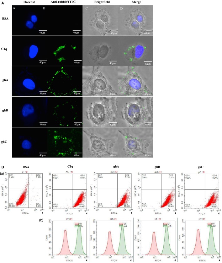
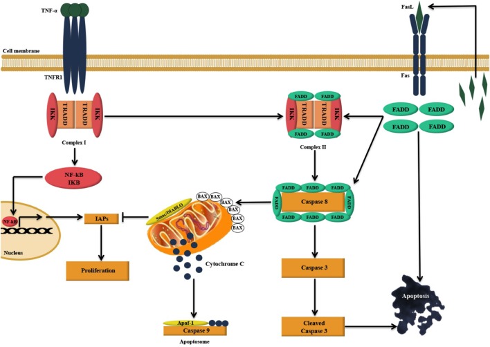
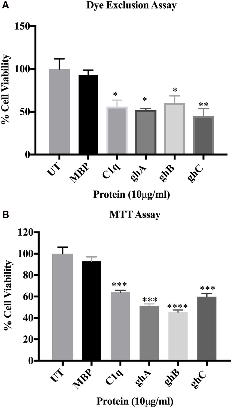
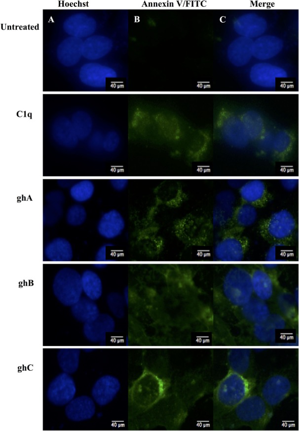
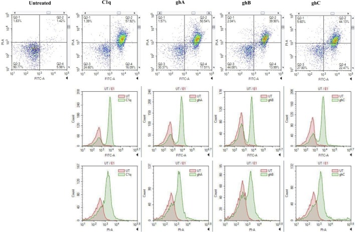
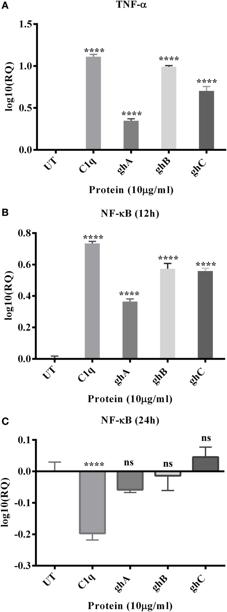
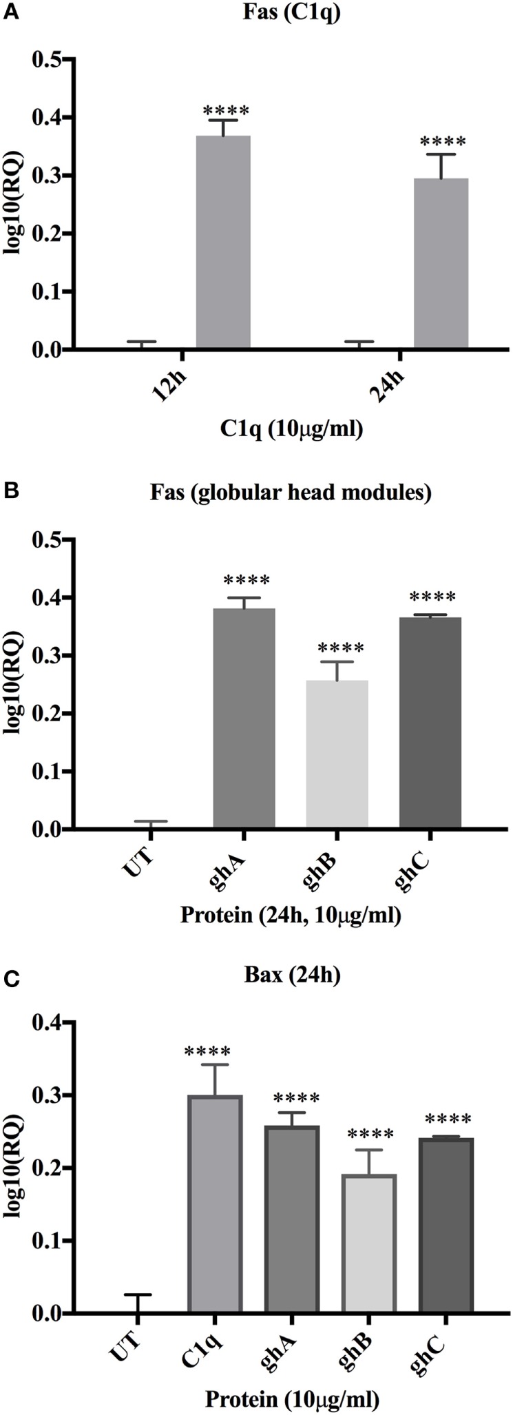
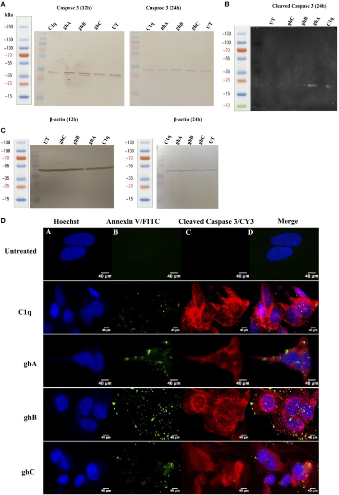
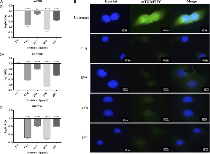
Complement protein C1q is the first recognition subcomponent of the complement classical pathway that plays a vital role in the clearance of immune complexes, pathogens, and apoptotic cells. C1q also has a homeostatic role involving immune and non-immune cells; these functions not necessarily involve complement activation. Recently, C1q has been shown to be expressed locally in the microenvironment of a range of human malignant tumors, where it can promote cancer cell adhesion, migration, and proliferation, without involving complement activation. C1q has been shown to be present in the ascitic fluid formed during ovarian cancers. In this study, we have examined the effects of human C1q and its globular domain on an ovarian cancer cell line, SKOV3. We show that C1q and the recombinant globular head modules induce apoptosis in SKOV3 cells in a time-dependent manner. C1q expression was not detectable in the SKOV3 cells. Exogenous treatment with C1q and globular head modules at the concentration of 10 µg/ml induced apoptosis in approximately 55% cells, as revealed by immunofluorescence microscopy and FACS. The qPCR and caspase analysis suggested that C1q and globular head modules activated tumor necrosis factor (TNF)-α and upregulated Fas. The genes of mammalian target of rapamycin (mTOR), RICTOR, and RAPTOR survival pathways, which are often overexpressed in majority of the cancers, were significantly downregulated within few hours of the treatment of SKOV3 cells with C1q and globular head modules. In conclusion, C1q, its globular domain, induced apoptosis in an ovarian cancer cell line SKOV3 TNF-α induced apoptosis pathway involving upregulation of Bax and Fas. This study highlights a potentially protective role of C1q in certain cancers.

摘要

补体蛋白C1q是补体经典途径的首个识别亚成分,在免疫复合物、病原体和凋亡细胞的清除中起关键作用。C1q在免疫和非免疫细胞中也具有稳态作用;这些功能不一定涉及补体激活。最近研究表明,C1q在一系列人类恶性肿瘤的微环境中局部表达,在不涉及补体激活的情况下,它可促进癌细胞的黏附、迁移和增殖。研究表明,C1q存在于卵巢癌形成的腹水中。在本研究中,我们检测了人C1q及其球状结构域对卵巢癌细胞系SKOV3的影响。我们发现,C1q和重组球状头部模块以时间依赖性方式诱导SKOV3细胞凋亡。在SKOV3细胞中未检测到C1q表达。免疫荧光显微镜和流式细胞术显示,用浓度为10μg/ml的C1q和球状头部模块进行外源处理可诱导约55%的细胞凋亡。定量PCR和半胱天冬酶分析表明,C1q和球状头部模块激活了肿瘤坏死因子(TNF)-α并上调了Fas。在大多数癌症中常过度表达的雷帕霉素哺乳动物靶标(mTOR)、RICTOR和RAPTOR生存途径的基因,在用C1q和球状头部模块处理SKOV3细胞的数小时内显著下调。总之,C1q及其球状结构域通过涉及上调Bax和Fas的TNF-α诱导凋亡途径,在卵巢癌细胞系SKOV3中诱导凋亡。本研究突出了C1q在某些癌症中潜在的保护作用。

https://cdn.ncbi.nlm.nih.gov/pmc/blobs/3d2e/5174108/5032add4ee64/fimmu-07-00599-g009.jpg
https://cdn.ncbi.nlm.nih.gov/pmc/blobs/3d2e/5174108/0f114acd9244/fimmu-07-00599-g001.jpg
https://cdn.ncbi.nlm.nih.gov/pmc/blobs/3d2e/5174108/abcd66013b84/fimmu-07-00599-g002.jpg
https://cdn.ncbi.nlm.nih.gov/pmc/blobs/3d2e/5174108/e0ce872f4ffe/fimmu-07-00599-g003.jpg
https://cdn.ncbi.nlm.nih.gov/pmc/blobs/3d2e/5174108/69b3aacb87c5/fimmu-07-00599-g004.jpg
https://cdn.ncbi.nlm.nih.gov/pmc/blobs/3d2e/5174108/511c02f5cfcc/fimmu-07-00599-g005.jpg
https://cdn.ncbi.nlm.nih.gov/pmc/blobs/3d2e/5174108/7174b9f8198d/fimmu-07-00599-g006.jpg
https://cdn.ncbi.nlm.nih.gov/pmc/blobs/3d2e/5174108/06b282fdd173/fimmu-07-00599-g007.jpg
https://cdn.ncbi.nlm.nih.gov/pmc/blobs/3d2e/5174108/e91a66d6a8f2/fimmu-07-00599-g008.jpg
https://cdn.ncbi.nlm.nih.gov/pmc/blobs/3d2e/5174108/5032add4ee64/fimmu-07-00599-g009.jpg

相似文献

1
Human C1q Induces Apoptosis in an Ovarian Cancer Cell Line Tumor Necrosis Factor Pathway.人C1q通过肿瘤坏死因子途径诱导卵巢癌细胞系凋亡。
Front Immunol. 2016 Dec 21;7:599. doi: 10.3389/fimmu.2016.00599. eCollection 2016.
2
Complement Protein C1q Binds to Hyaluronic Acid in the Malignant Pleural Mesothelioma Microenvironment and Promotes Tumor Growth.补体蛋白C1q与恶性胸膜间皮瘤微环境中的透明质酸结合并促进肿瘤生长。
Front Immunol. 2017 Nov 20;8:1559. doi: 10.3389/fimmu.2017.01559. eCollection 2017.
3
Recent progress in the understanding of the structure-function relationships of the globular head regions of C1q.C1q球状头部区域结构-功能关系理解方面的最新进展。
Immunobiology. 2002 Sep;205(4-5):355-64. doi: 10.1078/0171-2985-00138.
4
Complement Protein C1q Interacts with DC-SIGN Its Globular Domain and Thus May Interfere with HIV-1 Transmission.补体蛋白C1q与DC-SIGN及其球状结构域相互作用,因此可能干扰HIV-1传播。
Front Immunol. 2016 Dec 22;7:600. doi: 10.3389/fimmu.2016.00600. eCollection 2016.
5
Direct binding of C1q to apoptotic cells and cell blebs induces complement activation.C1q与凋亡细胞及细胞小泡的直接结合会诱导补体激活。
Eur J Immunol. 2002 Jun;32(6):1726-36. doi: 10.1002/1521-4141(200206)32:6<1726::AID-IMMU1726>3.0.CO;2-R.
6
The non-classical functions of the classical complement pathway recognition subcomponent C1q.经典补体途径识别亚基 C1q 的非经典功能。
Immunol Lett. 2010 Jul 8;131(2):139-50. doi: 10.1016/j.imlet.2010.03.012. Epub 2010 Apr 8.
7
Specific inhibition of the classical complement pathway with an engineered single-chain Fv to C1q globular heads decreases complement activation by apoptotic cells.工程化单链 Fv 特异性抑制经典补体途径的 C1q 球形头部可减少凋亡细胞补体的激活。
Immunobiology. 2010 May;215(5):395-405. doi: 10.1016/j.imbio.2009.05.010. Epub 2009 Jul 7.
8
Analysis of the Interaction between Globular Head Modules of Human C1q and Its Candidate Receptor gC1qR.人C1q球状头部模块与其候选受体gC1qR之间的相互作用分析
Front Immunol. 2016 Dec 13;7:567. doi: 10.3389/fimmu.2016.00567. eCollection 2016.
9
Modular organization of proteins containing C1q-like globular domain.含C1q样球状结构域蛋白质的模块化组织
Immunopharmacology. 1999 May;42(1-3):15-21. doi: 10.1016/s0162-3109(99)00011-9.
10
Succinate dehydrogenase subunit B inhibits the AMPK-HIF-1α pathway in human ovarian cancer in vitro.琥珀酸脱氢酶亚基B在体外抑制人卵巢癌中的AMPK-HIF-1α通路。
J Ovarian Res. 2014 Dec 10;7:115. doi: 10.1186/s13048-014-0115-1.

引用本文的文献

1
KCC2 inhibition and neuronal hyperexcitability promote extrinsic apoptosis dependent upon C1q.钾氯共转运体2(KCC2)抑制和神经元过度兴奋促进依赖于C1q的外源性凋亡。
Front Mol Neurosci. 2025 Aug 18;18:1645428. doi: 10.3389/fnmol.2025.1645428. eCollection 2025.
2
C1q Macrophage-Tumor Cell Interaction Promoted Tumorigenesis via GPR17/PI3K/AKT Pathway Induced DNA Hypermethylation in Nasopharyngeal Carcinoma.C1q巨噬细胞-肿瘤细胞相互作用通过GPR17/PI3K/AKT途径诱导鼻咽癌DNA高甲基化促进肿瘤发生。
Adv Sci (Weinh). 2025 Apr 2:e2503434. doi: 10.1002/advs.202503434.
3
Adipocyte-derived CXCL10 in obesity promotes the migration and invasion of ovarian cancer cells.

本文引用的文献

1
Editorial: State-of-the-Art Research on C1q and the Classical Complement Pathway.社论:C1q与经典补体途径的前沿研究
Front Immunol. 2016 Oct 4;7:398. doi: 10.3389/fimmu.2016.00398. eCollection 2016.
2
C1q acts in the tumour microenvironment as a cancer-promoting factor independently of complement activation.C1q在肿瘤微环境中作为一种促癌因子发挥作用,与补体激活无关。
Nat Commun. 2016 Feb 1;7:10346. doi: 10.1038/ncomms10346.
3
Emerging and Novel Functions of Complement Protein C1q.补体蛋白C1q的新出现及新颖功能
肥胖状态下脂肪细胞衍生的CXCL10促进卵巢癌细胞的迁移和侵袭。
J Ovarian Res. 2024 Dec 19;17(1):245. doi: 10.1186/s13048-024-01568-0.
4
Causal relationship between complement and colorectal cancer: a drug target Mendelian randomization study.补体与结直肠癌之间的因果关系:一项药物靶点孟德尔随机化研究。
Front Genet. 2024 Jul 23;15:1403509. doi: 10.3389/fgene.2024.1403509. eCollection 2024.
5
Absolute Quantification of Pan-Cancer Plasma Proteomes Reveals Unique Signature in Multiple Myeloma.泛癌血浆蛋白质组的绝对定量揭示了多发性骨髓瘤的独特特征。
Cancers (Basel). 2023 Sep 28;15(19):4764. doi: 10.3390/cancers15194764.
6
Detection of Complement C1q B Chain Overexpression and Its Latent Molecular Mechanisms in Cervical Cancer Tissues Using Multiple Methods.多种方法检测宫颈癌组织中补体C1q B链过表达及其潜在分子机制
Int J Genomics. 2022 Oct 20;2022:8775330. doi: 10.1155/2022/8775330. eCollection 2022.
7
Prognostic and immune-related value of complement C1Q (C1QA, C1QB, and C1QC) in skin cutaneous melanoma.补体C1Q(C1QA、C1QB和C1QC)在皮肤黑色素瘤中的预后及免疫相关价值
Front Genet. 2022 Aug 30;13:940306. doi: 10.3389/fgene.2022.940306. eCollection 2022.
8
Bax Contributes to Retinal Ganglion Cell Dendritic Degeneration During Glaucoma.Bax 促进青光眼期间视网膜神经节细胞树突退化。
Mol Neurobiol. 2022 Mar;59(3):1366-1380. doi: 10.1007/s12035-021-02675-5. Epub 2022 Jan 5.
9
Modulation of the Complement System by Neoplastic Disease of the Central Nervous System.中枢神经系统肿瘤性疾病对补体系统的调节。
Front Immunol. 2021 Oct 4;12:689435. doi: 10.3389/fimmu.2021.689435. eCollection 2021.
10
The Complement System in Ovarian Cancer: An Underexplored Old Path.卵巢癌中的补体系统:一条未被充分探索的古老途径。
Cancers (Basel). 2021 Jul 28;13(15):3806. doi: 10.3390/cancers13153806.
Front Immunol. 2015 Jun 29;6:317. doi: 10.3389/fimmu.2015.00317. eCollection 2015.
4
Therapeutic antibodies: Discovery, design and deployment.治疗性抗体:发现、设计与应用。
Mol Immunol. 2015 Oct;67(2 Pt A):1-3. doi: 10.1016/j.molimm.2015.05.004. Epub 2015 May 16.
5
Phosphatidylserine exposure during apoptosis reflects bidirectional trafficking between plasma membrane and cytoplasm.细胞凋亡过程中磷脂酰丝氨酸的暴露反映了质膜和细胞质之间的双向转运。
Cell Death Differ. 2013 Jan;20(1):64-76. doi: 10.1038/cdd.2012.93. Epub 2012 Aug 3.
6
The C1q family of proteins: insights into the emerging non-traditional functions.C1q蛋白家族:对新兴非传统功能的见解
Front Immunol. 2012 Apr 5;3. doi: 10.3389/fimmu.2012.00052.
7
Antibody therapy of cancer.癌症的抗体治疗。
Nat Rev Cancer. 2012 Mar 22;12(4):278-87. doi: 10.1038/nrc3236.
8
The central role of initiator caspase-9 in apoptosis signal transduction and the regulation of its activation and activity on the apoptosome.起始半胱氨酸蛋白酶-9 在细胞凋亡信号转导中的核心作用及其在凋亡体上的激活和活性调节。
Exp Cell Res. 2012 Jul 1;318(11):1213-20. doi: 10.1016/j.yexcr.2012.02.013. Epub 2012 Feb 28.
9
The holo-apoptosome: activation of procaspase-9 and interactions with caspase-3.全凋亡体:前胱冬肽酶-9 的激活及与胱冬肽酶-3 的相互作用。
Structure. 2011 Aug 10;19(8):1084-96. doi: 10.1016/j.str.2011.07.001.
10
Complement activation by phospholipids: the interplay of factor H and C1q.磷脂对补体的激活:因子 H 和 C1q 的相互作用。
Protein Cell. 2010 Nov;1(11):1033-49. doi: 10.1007/s13238-010-0125-8. Epub 2010 Dec 10.