• 文献检索
  • 文档翻译
  • 深度研究
  • 学术资讯
  • Suppr Zotero 插件Zotero 插件
  • 邀请有礼
  • 套餐&价格
  • 历史记录
应用&插件
Suppr Zotero 插件Zotero 插件浏览器插件Mac 客户端Windows 客户端微信小程序
定价
高级版会员购买积分包购买API积分包
服务
文献检索文档翻译深度研究API 文档MCP 服务
关于我们
关于 Suppr公司介绍联系我们用户协议隐私条款
关注我们

Suppr 超能文献

核心技术专利:CN118964589B侵权必究
粤ICP备2023148730 号-1Suppr @ 2026

文献检索

告别复杂PubMed语法,用中文像聊天一样搜索,搜遍4000万医学文献。AI智能推荐,让科研检索更轻松。

立即免费搜索

文件翻译

保留排版,准确专业,支持PDF/Word/PPT等文件格式,支持 12+语言互译。

免费翻译文档

深度研究

AI帮你快速写综述,25分钟生成高质量综述,智能提取关键信息,辅助科研写作。

立即免费体验

乳腺病理学中的计算机图像分析

Computer-based image analysis in breast pathology.

作者信息

Gandomkar Ziba, Brennan Patrick C, Mello-Thoms Claudia

机构信息

Image Optimisation and Perception, Discipline of Medical Radiation Sciences, University of Sydney, Australia.

Image Optimisation and Perception, Discipline of Medical Radiation Sciences, University of Sydney, Australia; Department of Biomedical Informatics, University of Pittsburgh School of Medicine, Pittsburgh, Pennsylvania, USA.

出版信息

J Pathol Inform. 2016 Oct 21;7:43. doi: 10.4103/2153-3539.192814. eCollection 2016.

DOI:10.4103/2153-3539.192814
PMID:28066683
原文链接:https://pmc.ncbi.nlm.nih.gov/articles/PMC5100199/
Abstract

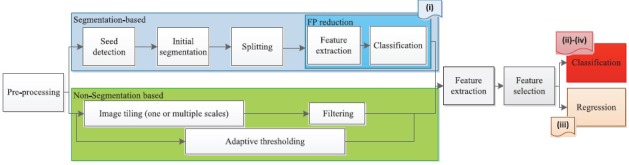
Whole slide imaging (WSI) has the potential to be utilized in telepathology, teleconsultation, quality assurance, clinical education, and digital image analysis to aid pathologists. In this paper, the potential added benefits of computer-assisted image analysis in breast pathology are reviewed and discussed. One of the major advantages of WSI systems is the possibility of doing computer-based image analysis on the digital slides. The purpose of computer-assisted analysis of breast virtual slides can be (i) segmentation of desired regions or objects such as diagnostically relevant areas, epithelial nuclei, lymphocyte cells, tubules, and mitotic figures, (ii) classification of breast slides based on breast cancer (BCa) grades, the invasive potential of tumors, or cancer subtypes, (iii) prognosis of BCa, or (iv) immunohistochemical quantification. While encouraging results have been achieved in this area, further progress is still required to make computer-based image analysis of breast virtual slides acceptable for clinical practice.

摘要

全玻片成像(WSI)有潜力应用于远程病理学、远程会诊、质量保证、临床教育以及数字图像分析,以辅助病理学家。本文回顾并讨论了计算机辅助图像分析在乳腺病理学中的潜在附加益处。WSI系统的主要优势之一是能够对数字玻片进行基于计算机的图像分析。乳腺虚拟玻片计算机辅助分析的目的可以是:(i)分割所需区域或对象,如诊断相关区域、上皮细胞核、淋巴细胞、小管和有丝分裂图像;(ii)根据乳腺癌(BCa)分级、肿瘤侵袭潜力或癌症亚型对乳腺玻片进行分类;(iii)预测BCa;或(iv)免疫组织化学定量分析。尽管该领域已取得令人鼓舞的成果,但仍需进一步进展,以使乳腺虚拟玻片的基于计算机的图像分析在临床实践中被接受。

https://cdn.ncbi.nlm.nih.gov/pmc/blobs/3984/5100199/70fd380dceb6/JPI-7-43-g001.jpg

相似文献

1
Computer-based image analysis in breast pathology.乳腺病理学中的计算机图像分析
J Pathol Inform. 2016 Oct 21;7:43. doi: 10.4103/2153-3539.192814. eCollection 2016.
2
Digital vs traditional: Are diagnostic accuracy rates similar for glass slides vs whole slide images in a non-gynaecological external quality assurance setting?数字与传统:在非妇科外部质量保证环境中,玻片与全切片图像的诊断准确率相似吗?
Cytopathology. 2018 Aug;29(4):326-334. doi: 10.1111/cyt.12552. Epub 2018 May 16.
3
Applications and challenges of digital pathology and whole slide imaging.数字病理学与全切片成像的应用及挑战
Biotech Histochem. 2015 Jul;90(5):341-7. doi: 10.3109/10520295.2015.1044566. Epub 2015 May 15.
4
Eye-movement study and human performance using telepathology virtual slides: implications for medical education and differences with experience.使用远程病理学虚拟切片的眼动研究与人类表现:对医学教育的启示及与经验的差异
Hum Pathol. 2006 Dec;37(12):1543-56. doi: 10.1016/j.humpath.2006.08.024.
5
Virtual slide telepathology for an academic teaching hospital surgical pathology quality assurance program.用于学术教学医院外科病理学质量保证计划的虚拟玻片远程病理学
Hum Pathol. 2009 Aug;40(8):1129-36. doi: 10.1016/j.humpath.2009.04.008. Epub 2009 Jun 21.
6
Utility of whole slide imaging and virtual microscopy in prostate pathology.全玻片成像和虚拟显微镜在前列腺病理学中的应用。
APMIS. 2012 Apr;120(4):298-304. doi: 10.1111/j.1600-0463.2011.02872.x.
7
Diagnosis of major cancer resection specimens with virtual slides: impact of a novel digital pathology workstation.使用虚拟切片对主要癌症切除标本进行诊断:新型数字病理工作站的影响
Hum Pathol. 2014 Oct;45(10):2101-6. doi: 10.1016/j.humpath.2014.06.017. Epub 2014 Jul 2.
8
Exploring virtual reality technology and the Oculus Rift for the examination of digital pathology slides.探索虚拟现实技术及Oculus Rift用于数字病理切片检查。
J Pathol Inform. 2016 May 4;7:22. doi: 10.4103/2153-3539.181766. eCollection 2016.
9
New Trends of Emerging Technologies in Digital Pathology.数字病理学中新兴技术的新趋势
Pathobiology. 2016;83(2-3):61-9. doi: 10.1159/000443482. Epub 2016 Apr 26.
10
Virtual slide telepathology enables an innovative telehealth rapid breast care clinic.虚拟玻片远程病理学助力创新的远程医疗快速乳腺护理诊所。
Hum Pathol. 2009 Aug;40(8):1082-91. doi: 10.1016/j.humpath.2009.04.005. Epub 2009 Jun 23.

引用本文的文献

1
Higher density of compact B cell clusters in invasive front may contribute to better prognosis in pancreatic ductal adenocarcinoma.侵袭前沿致密B细胞簇的更高密度可能有助于改善胰腺导管腺癌的预后。
Discov Oncol. 2025 Apr 17;16(1):555. doi: 10.1007/s12672-025-02260-5.
2
Semi-automated analysis of HER2 immunohistochemistry in invasive breast carcinoma using whole slide images: utility for interpretation in clinical practice.使用全切片图像对半自动化分析浸润性乳腺癌的 HER2 免疫组化:在临床实践中的解释效用。
Pathol Oncol Res. 2024 Aug 29;30:1611826. doi: 10.3389/pore.2024.1611826. eCollection 2024.
3
Revolutionizing Pathology with Artificial Intelligence: Innovations in Immunohistochemistry.

本文引用的文献

1
Breast histopathology image segmentation using spatio-colour-texture based graph partition method.基于空间颜色纹理的图划分方法的乳腺组织病理学图像分割
J Microsc. 2016 Jun;262(3):260-73. doi: 10.1111/jmi.12361. Epub 2015 Dec 28.
2
Triaging Diagnostically Relevant Regions from Pathology Whole Slides of Breast Cancer: A Texture Based Approach.从乳腺癌全切片病理中筛选有诊断意义的区域:一种基于纹理的方法。
IEEE Trans Med Imaging. 2016 Jan;35(1):307-15. doi: 10.1109/TMI.2015.2470529. Epub 2015 Aug 20.
3
A Model based Survey of Colour Deconvolution in Diagnostic Brightfield Microscopy: Error Estimation and Spectral Consideration.
人工智能引领病理学变革:免疫组织化学的创新
J Pers Med. 2024 Jun 27;14(7):693. doi: 10.3390/jpm14070693.
4
Augmented interpretation of HER2, ER, and PR in breast cancer by artificial intelligence analyzer: enhancing interobserver agreement through a reader study of 201 cases.人工智能分析器增强乳腺癌中 HER2、ER 和 PR 的解读:通过对 201 例病例的读片研究提高观察者间一致性。
Breast Cancer Res. 2024 Feb 23;26(1):31. doi: 10.1186/s13058-024-01784-y.
5
An interpretable decision-support model for breast cancer diagnosis using histopathology images.一种使用组织病理学图像进行乳腺癌诊断的可解释决策支持模型。
J Pathol Inform. 2023 Jun 13;14:100319. doi: 10.1016/j.jpi.2023.100319. eCollection 2023.
6
Analysis of Image Feature Characteristics for Automated Scoring of HER2 in Histology Slides.用于组织学切片中HER2自动评分的图像特征分析
J Imaging. 2019 Mar 10;5(3):35. doi: 10.3390/jimaging5030035.
7
Using Digital Pathology to Understand Epithelial Characteristics of Benign Breast Disease among Women Undergoing Diagnostic Image-Guided Breast Biopsy.利用数字病理学了解接受影像引导乳腺活检的女性良性乳腺疾病的上皮特征。
Cancer Prev Res (Phila). 2019 Dec;12(12):861-870. doi: 10.1158/1940-6207.CAPR-19-0120. Epub 2019 Oct 23.
8
A deep learning algorithm for one-step contour aware nuclei segmentation of histopathology images.一种用于病理图像一步轮廓感知细胞核分割的深度学习算法。
Med Biol Eng Comput. 2019 Sep;57(9):2027-2043. doi: 10.1007/s11517-019-02008-8. Epub 2019 Jul 26.
9
Computer-Assisted Nuclear Atypia Scoring of Breast Cancer: a Preliminary Study.计算机辅助乳腺癌核异型性评分:一项初步研究。
J Digit Imaging. 2019 Oct;32(5):702-712. doi: 10.1007/s10278-019-00181-8.
10
Quantitative Characterization of CD8+ T Cell Clustering and Spatial Heterogeneity in Solid Tumors.实体瘤中CD8 + T细胞聚集和空间异质性的定量表征
Front Oncol. 2019 Jan 7;8:649. doi: 10.3389/fonc.2018.00649. eCollection 2018.
基于模型的诊断明场显微镜中颜色反卷积的调查:误差估计与光谱考量
Sci Rep. 2015 Jul 30;5:12096. doi: 10.1038/srep12096.
4
Computer Aided Semi-Automated Evaluation of HER2 Immunodetection--A Robust Solution for Supporting the Accuracy of Anti HER2 Therapy.人表皮生长因子受体2免疫检测的计算机辅助半自动评估——支持抗人表皮生长因子受体2治疗准确性的可靠解决方案
Pathol Oncol Res. 2015 Sep;21(4):1005-11. doi: 10.1007/s12253-015-9927-6. Epub 2015 Mar 19.
5
Assessment of algorithms for mitosis detection in breast cancer histopathology images.乳腺癌组织病理学图像有丝分裂检测算法的评估。
Med Image Anal. 2015 Feb;20(1):237-48. doi: 10.1016/j.media.2014.11.010. Epub 2014 Nov 29.
6
Towards semantic-driven high-content image analysis: an operational instantiation for mitosis detection in digital histopathology.面向语义驱动的高内涵图像分析:数字组织病理学中用于有丝分裂检测的操作实例。
Comput Med Imaging Graph. 2015 Jun;42:2-15. doi: 10.1016/j.compmedimag.2014.09.004. Epub 2014 Oct 2.
7
Effects of tissue decalcification on the quantification of breast cancer biomarkers by digital image analysis.组织脱钙对通过数字图像分析定量乳腺癌生物标志物的影响。
Diagn Pathol. 2014 Nov 25;9:213. doi: 10.1186/s13000-014-0213-9.
8
A nonlinear mapping approach to stain normalization in digital histopathology images using image-specific color deconvolution.一种使用特定图像颜色反卷积对数字组织病理学图像进行染色归一化的非线性映射方法。
IEEE Trans Biomed Eng. 2014 Jun;61(6):1729-38. doi: 10.1109/TBME.2014.2303294.
9
Mitosis detection in breast cancer histology images with deep neural networks.利用深度神经网络检测乳腺癌组织学图像中的有丝分裂
Med Image Comput Comput Assist Interv. 2013;16(Pt 2):411-8. doi: 10.1007/978-3-642-40763-5_51.
10
Multi-label fast marching and seeded watershed segmentation methods for diagnosis of breast cancer cytology.用于乳腺癌细胞学诊断的多标签快速行进和种子区域生长分割方法
Annu Int Conf IEEE Eng Med Biol Soc. 2013;2013:7368-71. doi: 10.1109/EMBC.2013.6611260.