• 文献检索
  • 文档翻译
  • 深度研究
  • 学术资讯
  • Suppr Zotero 插件Zotero 插件
  • 邀请有礼
  • 套餐&价格
  • 历史记录
应用&插件
Suppr Zotero 插件Zotero 插件浏览器插件Mac 客户端Windows 客户端微信小程序
定价
高级版会员购买积分包购买API积分包
服务
文献检索文档翻译深度研究API 文档MCP 服务
关于我们
关于 Suppr公司介绍联系我们用户协议隐私条款
关注我们

Suppr 超能文献

核心技术专利:CN118964589B侵权必究
粤ICP备2023148730 号-1Suppr @ 2026

文献检索

告别复杂PubMed语法,用中文像聊天一样搜索,搜遍4000万医学文献。AI智能推荐,让科研检索更轻松。

立即免费搜索

文件翻译

保留排版,准确专业,支持PDF/Word/PPT等文件格式,支持 12+语言互译。

免费翻译文档

深度研究

AI帮你快速写综述,25分钟生成高质量综述,智能提取关键信息,辅助科研写作。

立即免费体验

利用边缘密度成像进行连接组学图谱绘制可区分儿童轻度创伤性脑损伤与正常发育对照:概念验证。

Connectome mapping with edge density imaging differentiates pediatric mild traumatic brain injury from typically developing controls: proof of concept.

机构信息

Neural Connectivity Laboratory, Department of Radiology and Biomedical Imaging, University of California, San Francisco, 185 Berry St., Suite 350, San Francisco, CA, 94158, USA.

Mallinckrodt Institute of Radiology, Division of Neuroradiology, Washington University in St. Louis, St. Louis, MO, USA.

出版信息

Pediatr Radiol. 2020 Oct;50(11):1594-1601. doi: 10.1007/s00247-020-04743-9. Epub 2020 Jun 30.

DOI:10.1007/s00247-020-04743-9
PMID:32607611
原文链接:https://pmc.ncbi.nlm.nih.gov/articles/PMC7501221/
Abstract

BACKGROUND

Although acute neurologic impairment might be transient, other long-term effects can be observed with mild traumatic brain injury. However, when pediatric patients with mild traumatic brain injury present for medical care, conventional imaging with CT and MR imaging often does not reveal abnormalities.

OBJECTIVE

To determine whether edge density imaging can separate pediatric mild traumatic brain injury from typically developing controls.

MATERIALS AND METHODS

Subjects were recruited as part of the "Therapeutic Resources for Attention Improvement using Neuroimaging in Traumatic Brain Injury" (TRAIN-TBI) study. We included 24 adolescents (χ=14.1 years of age, σ=1.6 years, range 10-16 years), 14 with mild traumatic brain injury (TBI) and 10 typically developing controls. Neurocognitive assessments included the pediatric version of the California Verbal Learning Test (CVLT) and the Attention Network Task (ANT). Diffusion MR imaging was acquired on a 3-tesla (T) scanner. Edge density images were computed utilizing fiber tractography. Principal component analysis (PCA) and support vector machines (SVM) were used in an exploratory analysis to separate mild TBI and control groups. The diagnostic accuracy of edge density imaging, neurocognitive tests, and fractional anisotropy (FA) from diffusion tensor imaging (DTI) was computed with two-sample t-tests and receiver operating characteristic (ROC) metrics.

RESULTS

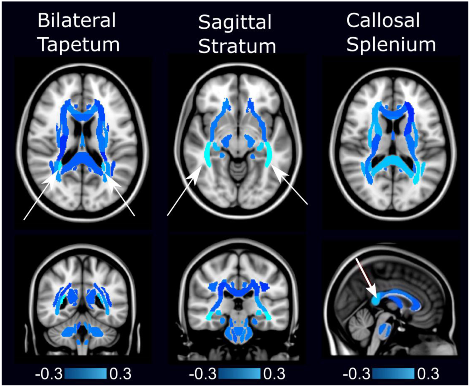
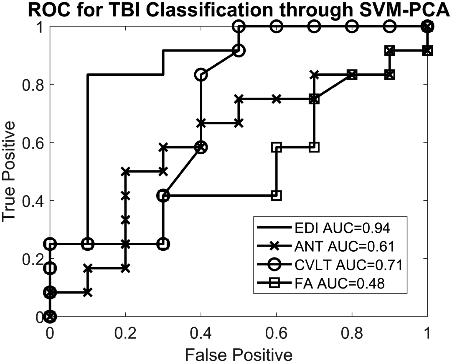
Support vector machine-principal component analysis of edge density imaging maps identified three white matter regions distinguishing pediatric mild TBI from controls. The bilateral tapetum, sagittal stratum, and callosal splenium identified mild TBI subjects with sensitivity of 79% and specificity of 100%. Accuracy from the area under the ROC curve (AUC) was 94%. Neurocognitive testing provided an AUC of 61% (CVLT) and 71% (ANT). Fractional anisotropy yielded an AUC of 48%.

CONCLUSION

In this proof-of-concept study, we show that edge density imaging is a new form of connectome mapping that provides better diagnostic delineation between pediatric mild TBI and healthy controls than DTI or neurocognitive assessments of memory or attention.

摘要

背景

尽管急性神经功能障碍可能是短暂的,但轻度创伤性脑损伤也可能出现其他长期影响。然而,当儿科轻度创伤性脑损伤患者接受医疗护理时,常规的 CT 和磁共振成像通常不会发现异常。

目的

确定边缘密度成像是否可以区分儿科轻度创伤性脑损伤和正常发育的对照组。

材料和方法

研究对象是“使用神经影像学治疗创伤性脑损伤注意力改善的治疗资源”(TRAIN-TBI)研究的一部分。我们纳入了 24 名青少年(χ=14.1 岁,σ=1.6 岁,范围 10-16 岁),其中 14 名患有轻度创伤性脑损伤(TBI),10 名正常发育的对照组。神经认知评估包括儿童版加利福尼亚语言学习测试(CVLT)和注意力网络测试(ANT)。扩散磁共振成像在 3 特斯拉(T)扫描仪上采集。利用纤维束追踪技术计算边缘密度图像。主成分分析(PCA)和支持向量机(SVM)用于探索性分析,以区分轻度 TBI 和对照组。使用两样本 t 检验和接收者操作特征(ROC)指标计算边缘密度成像、神经认知测试和来自扩散张量成像(DTI)的各向异性分数(FA)的诊断准确性。

结果

边缘密度成像图谱的支持向量机-主成分分析确定了三个区分儿科轻度 TBI 和对照组的白质区域。双侧 tapetum、矢状层和胼胝体压部可识别轻度 TBI 患者,敏感性为 79%,特异性为 100%。ROC 曲线下面积(AUC)的准确性为 94%。神经认知测试的 AUC 分别为 61%(CVLT)和 71%(ANT)。各向异性分数的 AUC 为 48%。

结论

在这项概念验证研究中,我们表明边缘密度成像作为一种新的连接组学映射形式,与 DTI 或记忆或注意力的神经认知评估相比,提供了更好的儿科轻度 TBI 和健康对照组之间的诊断区分。

https://cdn.ncbi.nlm.nih.gov/pmc/blobs/eaea/7501221/5a7367808f29/nihms-1608523-f0003.jpg
https://cdn.ncbi.nlm.nih.gov/pmc/blobs/eaea/7501221/bb3eff243ad0/nihms-1608523-f0001.jpg
https://cdn.ncbi.nlm.nih.gov/pmc/blobs/eaea/7501221/c2ca87b11242/nihms-1608523-f0002.jpg
https://cdn.ncbi.nlm.nih.gov/pmc/blobs/eaea/7501221/5a7367808f29/nihms-1608523-f0003.jpg
https://cdn.ncbi.nlm.nih.gov/pmc/blobs/eaea/7501221/bb3eff243ad0/nihms-1608523-f0001.jpg
https://cdn.ncbi.nlm.nih.gov/pmc/blobs/eaea/7501221/c2ca87b11242/nihms-1608523-f0002.jpg
https://cdn.ncbi.nlm.nih.gov/pmc/blobs/eaea/7501221/5a7367808f29/nihms-1608523-f0003.jpg

相似文献

1
Connectome mapping with edge density imaging differentiates pediatric mild traumatic brain injury from typically developing controls: proof of concept.利用边缘密度成像进行连接组学图谱绘制可区分儿童轻度创伤性脑损伤与正常发育对照:概念验证。
Pediatr Radiol. 2020 Oct;50(11):1594-1601. doi: 10.1007/s00247-020-04743-9. Epub 2020 Jun 30.
2
Relevance of neuroimaging for neurocognitive and behavioral outcome after pediatric traumatic brain injury.神经影像学在儿童创伤性脑损伤后神经认知和行为结果中的相关性。
Brain Imaging Behav. 2018 Feb;12(1):29-43. doi: 10.1007/s11682-017-9673-3.
3
White Matter Connectome Edge Density in Children with Autism Spectrum Disorders: Potential Imaging Biomarkers Using Machine-Learning Models.自闭症谱系障碍儿童的白质连接体边缘密度:基于机器学习模型的潜在影像生物标志物。
Brain Connect. 2019 Mar;9(2):209-220. doi: 10.1089/brain.2018.0658.
4
Acute pediatric traumatic brain injury severity predicts long-term verbal memory performance through suppression by white matter integrity on diffusion tensor imaging.急性儿科创伤性脑损伤严重程度通过弥散张量成像中的白质完整性抑制预测长期言语记忆表现。
Brain Imaging Behav. 2020 Oct;14(5):1626-1637. doi: 10.1007/s11682-019-00093-9.
5
Use of Support Vector Machines Approach via ComBat Harmonized Diffusion Tensor Imaging for the Diagnosis and Prognosis of Mild Traumatic Brain Injury: A CENTER-TBI Study.基于平衡化算法的支持向量机在轻度创伤性脑损伤诊断和预后中的弥散张量成像研究:CENTER-TBI 研究。
J Neurotrauma. 2023 Jul;40(13-14):1317-1338. doi: 10.1089/neu.2022.0365. Epub 2023 May 18.
6
Utility of diffusion tensor imaging in the acute stage of mild to moderate traumatic brain injury for detecting white matter lesions and predicting long-term cognitive function in adults.弥散张量成像在成人轻中度创伤性脑损伤急性期检测脑白质病变及预测远期认知功能中的应用。
J Neurosurg. 2011 Jul;115(1):130-9. doi: 10.3171/2011.2.JNS101547. Epub 2011 Mar 18.
7
White matter integrity and cognition in chronic traumatic brain injury: a diffusion tensor imaging study.慢性创伤性脑损伤中的白质完整性与认知:一项扩散张量成像研究
Brain. 2007 Oct;130(Pt 10):2508-19. doi: 10.1093/brain/awm216. Epub 2007 Sep 14.
8
Common Patterns of Regional Brain Injury Detectable by Diffusion Tensor Imaging in Otherwise Normal-Appearing White Matter in Patients with Early Moderate to Severe Traumatic Brain Injury.早期中重度创伤性脑损伤患者,弥散张量成像可发现正常外观的脑白质中存在的常见区域性脑损伤模式。
J Neurotrauma. 2018 Mar 1;35(5):739-749. doi: 10.1089/neu.2016.4944. Epub 2018 Feb 9.
9
Changes in Apparent Fiber Density and Track-Weighted Imaging Metrics in White Matter following Experimental Traumatic Brain Injury.实验性创伤性脑损伤后白质中表观纤维密度和轨迹加权成像指标的变化
J Neurotrauma. 2017 Jul 1;34(13):2109-2118. doi: 10.1089/neu.2016.4730. Epub 2017 Apr 13.
10
Statistical machine learning to identify traumatic brain injury (TBI) from structural disconnections of white matter networks.利用统计机器学习从白质网络的结构断开中识别创伤性脑损伤(TBI)。
Neuroimage. 2016 Apr 1;129:247-259. doi: 10.1016/j.neuroimage.2016.01.056. Epub 2016 Jan 28.

引用本文的文献

1
Classification accuracy of structural and functional connectomes across different depressive phenotypes.不同抑郁表型的结构和功能连接组的分类准确率。
Imaging Neurosci (Camb). 2024 Jan 17;2. doi: 10.1162/imag_a_00064. eCollection 2024.
2
Artificial Intelligence in Pediatric Emergency Medicine: Applications, Challenges, and Future Perspectives.人工智能在儿科急诊医学中的应用、挑战及未来展望
Biomedicines. 2024 May 30;12(6):1220. doi: 10.3390/biomedicines12061220.
3
MaPPeRTrac: A Massively Parallel, Portable, and Reproducible Tractography Pipeline.

本文引用的文献

1
White Matter Connectome Edge Density in Children with Autism Spectrum Disorders: Potential Imaging Biomarkers Using Machine-Learning Models.自闭症谱系障碍儿童的白质连接体边缘密度:基于机器学习模型的潜在影像生物标志物。
Brain Connect. 2019 Mar;9(2):209-220. doi: 10.1089/brain.2018.0658.
2
Memory and the hippocampal formation following pediatric traumatic brain injury.儿童外伤性脑损伤后的记忆和海马结构。
Brain Behav. 2017 Nov 2;7(12):e00832. doi: 10.1002/brb3.832. eCollection 2017 Dec.
3
Prognostic value of corpus callosum injuries in severe head trauma.
MaPPeRTrac:一个大规模并行、可移植和可重复的轨迹追踪管道。
Neuroinformatics. 2024 Apr;22(2):177-191. doi: 10.1007/s12021-024-09650-0. Epub 2024 Mar 6.
4
Changes in the Networks of Attention across the Lifespan: A Graphical Meta-Analysis.一生中注意力网络的变化:一项图形荟萃分析。
J Intell. 2024 Feb 10;12(2):19. doi: 10.3390/jintelligence12020019.
5
Altered longitudinal structural connectome in paediatric mild traumatic brain injury: an Advancing Concussion Assessment in Paediatrics study.小儿轻度创伤性脑损伤中纵向结构连接组的改变:一项小儿脑震荡评估进展研究
Brain Commun. 2023 May 31;5(3):fcad173. doi: 10.1093/braincomms/fcad173. eCollection 2023.
6
Edge Density Imaging Identifies White Matter Biomarkers of Late-Life Obesity and Cognition.边缘密度成像可识别与晚年肥胖和认知相关的脑白质生物标志物。
Aging Dis. 2024 Aug 1;15(4):1899-1912. doi: 10.14336/AD.2022.1210.
7
Abnormal structural and functional network topological properties associated with left prefrontal, parietal, and occipital cortices significantly predict childhood TBI-related attention deficits: A semi-supervised deep learning study.与左前额叶、顶叶和枕叶皮质相关的异常结构和功能网络拓扑特性显著预测儿童创伤性脑损伤相关的注意力缺陷:一项半监督深度学习研究。
Front Neurosci. 2023 Mar 2;17:1128646. doi: 10.3389/fnins.2023.1128646. eCollection 2023.
8
Predicting sex, age, general cognition and mental health with machine learning on brain structural connectomes.基于脑结构连接组学的机器学习预测性别、年龄、一般认知和心理健康。
Hum Brain Mapp. 2023 Apr 1;44(5):1913-1933. doi: 10.1002/hbm.26182. Epub 2022 Dec 21.
9
Safeguarding Athletes Against Head Injuries Through Advances in Technology: A Scoping Review of the Uses of Machine Learning in the Management of Sports-Related Concussion.通过技术进步保护运动员免受头部损伤:机器学习在运动相关脑震荡管理中的应用范围综述
Front Sports Act Living. 2022 Apr 20;4:837643. doi: 10.3389/fspor.2022.837643. eCollection 2022.
10
The role of artificial intelligence in paediatric neuroradiology.人工智能在儿科神经放射学中的作用。
Pediatr Radiol. 2022 Oct;52(11):2159-2172. doi: 10.1007/s00247-022-05322-w. Epub 2022 Mar 26.
严重颅脑创伤中胼胝体损伤的预后价值
Acta Neurochir (Wien). 2017 Jan;159(1):25-32. doi: 10.1007/s00701-016-3000-4. Epub 2016 Oct 31.
4
Neurocognitive predictors of posttraumatic stress disorder symptoms in children 6 months after traumatic brain injury: A prospective study.创伤性脑损伤6个月后儿童创伤后应激障碍症状的神经认知预测因素:一项前瞻性研究。
Neuropsychology. 2017 Jan;31(1):84-92. doi: 10.1037/neu0000305. Epub 2016 Sep 12.
5
Periventricular White Matter Is a Nexus for Network Connectivity in the Human Brain.脑白质深部是人类大脑网络连接的枢纽。
Brain Connect. 2016 Sep;6(7):548-57. doi: 10.1089/brain.2016.0431.
6
Neuroimaging Wisely.明智地运用神经影像学。
AJNR Am J Neuroradiol. 2016 Dec;37(12):2182-2188. doi: 10.3174/ajnr.A4821. Epub 2016 Jun 9.
7
Epidemiology of Global Pediatric Traumatic Brain Injury: Qualitative Review.全球儿童创伤性脑损伤的流行病学:定性综述
World Neurosurg. 2016 Jul;91:497-509.e1. doi: 10.1016/j.wneu.2016.03.045. Epub 2016 Mar 25.
8
Sleep Disorders Associated With Mild Traumatic Brain Injury Using Sport Concussion Assessment Tool 3.使用运动性脑震荡评估工具3评估与轻度创伤性脑损伤相关的睡眠障碍
Pediatr Neurol. 2016 Apr;57:46-50.e1. doi: 10.1016/j.pediatrneurol.2015.12.019. Epub 2015 Dec 29.
9
White matter alterations in youth with acute mild traumatic brain injury.急性轻度创伤性脑损伤青年的白质改变
J Pediatr Rehabil Med. 2015;8(4):285-96. doi: 10.3233/PRM-150347.
10
The incidence of concussion in youth sports: a systematic review and meta-analysis.青少年运动中脑震荡的发生率:系统评价和荟萃分析。
Br J Sports Med. 2016 Mar;50(5):292-7. doi: 10.1136/bjsports-2015-094978. Epub 2015 Nov 30.